3E9R | pdb_00003e9r

Crystal structure of purine nucleoside phosphorylase from Schistosoma mansoni in complex with adenine


Experimental Data Snapshot

  • Method: X-RAY DIFFRACTION
  • Resolution: 1.85 Å
  • R-Value Free: 
    0.201 (Depositor), 0.201 (DCC) 
  • R-Value Work: 
    0.162 (Depositor), 0.161 (DCC) 
  • R-Value Observed: 
    0.164 (Depositor) 

Starting Model: experimental
View more details

wwPDB Validation 3D Report Full Report

Validation slider image for 3E9R

This is version 1.3 of the entry. See complete history

Literature

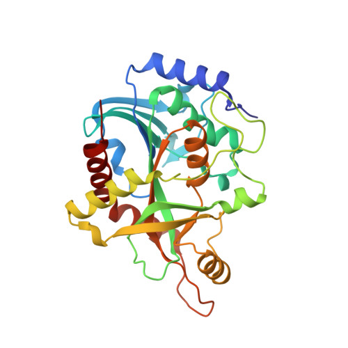
Adenosine binding to low-molecular-weight purine nucleoside phosphorylase: the structural basis for recognition based on its complex with the enzyme from Schistosoma mansoni.

Pereira, H.M.Rezende, M.M.Castilho, M.S.Oliva, G.Garratt, R.C.

(2010) Acta Crystallogr D Biol Crystallogr 66: 73-79

  • DOI: https://doi.org/10.1107/S0907444909045715
  • Primary Citation Related Structures: 
    3E9R, 3F8W, 3FAZ, 3FNQ

  • PubMed Abstract: 

    Schistosomes are unable to synthesize purines de novo and depend exclusively on the salvage pathway for their purine requirements. It has been suggested that blockage of this pathway could lead to parasite death. The enzyme purine nucleoside phosphorylase (PNP) is one of its key components and molecules designed to inhibit the low-molecular-weight (LMW) PNPs, which include both the human and schistosome enzymes, are typically analogues of the natural substrates inosine and guanosine. Here, it is shown that adenosine both binds to Schistosoma mansoni PNP and behaves as a weak micromolar inhibitor of inosine phosphorolysis. Furthermore, the first crystal structures of complexes of an LMW PNP with adenosine and adenine are reported, together with those with inosine and hypoxanthine. These are used to propose a structural explanation for the selective binding of adenosine to some LMW PNPs but not to others. The results indicate that transition-state analogues based on adenosine or other 6-amino nucleosides should not be discounted as potential starting points for alternative inhibitors.


  • Organizational Affiliation
    • Instituto de Física de São Carlos, Universidade de São Paulo, Avenida Trabalhador Sãocarlense 400, São Carlos, SP 13560-970, Brazil. hmuniz.pereira@gmail.com

Macromolecule Content 

  • Total Structure Weight: 94.46 kDa 
  • Atom Count: 7,206 
  • Modeled Residue Count: 842 
  • Deposited Residue Count: 861 
  • Unique protein chains: 1

Macromolecules

Find similar proteins by:|  3D Structure
Entity ID: 1
MoleculeChains  Sequence LengthOrganismDetailsImage
Purine-nucleoside phosphorylase
A, B, C
287Schistosoma mansoniMutation(s): 0 
Gene Names: SmPNP
EC: 2.4.2.1
UniProt
Find proteins for Q9BMI9 (Schistosoma mansoni)
Explore Q9BMI9 
Go to UniProtKB:  Q9BMI9
Entity Groups
Sequence Clusters30% Identity50% Identity70% Identity90% Identity95% Identity100% Identity
UniProt GroupQ9BMI9
Sequence Annotations
Expand
Reference Sequence

Small Molecules

Ligands 4 Unique
IDChains Name / Formula / InChI Key2D Diagram3D Interactions
ADE

Query on ADE


Download:Ideal Coordinates CCD File
H [auth B]ADENINE
C5 H5 N5
GFFGJBXGBJISGV-UHFFFAOYSA-N
SO4

Query on SO4


Download:Ideal Coordinates CCD File
D [auth A],
G [auth A],
I [auth B],
K [auth C]
SULFATE ION
O4 S
QAOWNCQODCNURD-UHFFFAOYSA-L
DMS

Query on DMS


Download:Ideal Coordinates CCD File
F [auth A],
J [auth B],
M [auth C]
DIMETHYL SULFOXIDE
C2 H6 O S
IAZDPXIOMUYVGZ-UHFFFAOYSA-N
ACT

Query on ACT


Download:Ideal Coordinates CCD File
E [auth A],
L [auth C]
ACETATE ION
C2 H3 O2
QTBSBXVTEAMEQO-UHFFFAOYSA-M

Experimental Data & Validation

Experimental Data

  • Method: X-RAY DIFFRACTION
  • Resolution: 1.85 Å
  • R-Value Free:  0.201 (Depositor), 0.201 (DCC) 
  • R-Value Work:  0.162 (Depositor), 0.161 (DCC) 
  • R-Value Observed: 0.164 (Depositor) 
Space Group: P 21 21 21
Unit Cell:
Length ( Å )Angle ( ˚ )
a = 48.989α = 90
b = 119.995β = 90
c = 130.982γ = 90
Software Package:
Software NamePurpose
MOSFLMdata reduction
SCALAdata scaling
MOLREPphasing
PHENIXrefinement
PDB_EXTRACTdata extraction
MAR345data collection

Structure Validation

View Full Validation Report


Entry History 

Deposition Data

Revision History  (Full details and data files)

  • Version 1.0: 2009-09-01
    Type: Initial release
  • Version 1.1: 2011-07-13
    Changes: Version format compliance
  • Version 1.2: 2017-10-25
    Changes: Refinement description
  • Version 1.3: 2023-08-30
    Changes: Data collection, Database references, Derived calculations, Refinement description