Crystal structure of the Homo sapiens kynureninase-3-hydroxyhippuric acid inhibitor complex: insights into the molecular basis of kynureninase substrate specificity.
Lima, S., Kumar, S., Gawandi, V., Momany, C., Phillips, R.S.(2009) J Med Chem 52: 389-396
- PubMed: 19143568 Search on PubMed
- DOI: https://doi.org/10.1021/jm8010806
- Primary Citation Related Structures:
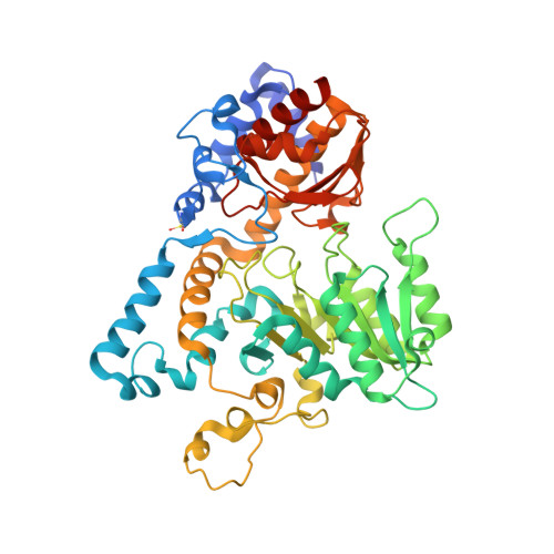
3E9K - PubMed Abstract:
Homo sapiens kynureninase is a pyridoxal-5'-phosphate dependent enzyme that catalyzes the hydrolytic cleavage of 3-hydroxykynurenine to yield 3-hydroxyanthranilate and L-alanine as part of the tryptophan catabolic pathway leading to the de novo biosynthesis of NAD(+). This pathway results in quinolinate, an excitotoxin that is an NMDA receptor agonist. High levels of quinolinate have been correlated with the etiology of neurodegenerative disorders such as AIDS-related dementia and Alzheimer's disease. We have synthesized a novel kynureninase inhibitor, 3-hydroxyhippurate, cocrystallized it with human kynureninase, and solved the atomic structure. On the basis of an analysis of the complex, we designed a series of His-102, Ser-332, and Asn-333 mutants. The H102W/N333T and H102W/S332G/N333T mutants showed complete reversal of substrate specificity between 3-hydroxykynurenine and L-kynurenine, thus defining the primary residues contributing to substrate specificity in kynureninases.
- Department of Biochemistry and Molecular Biology, University of Georgia, Athens, Georgia 30602, USA.
Organizational Affiliation: