3E53 | pdb_00003e53

Crystal structure of N-terminal domain of a Fatty Acyl AMP Ligase FAAL28 from Mycobacterium tuberculosis


Experimental Data Snapshot

  • Method: X-RAY DIFFRACTION
  • Resolution: 2.35 Å
  • R-Value Free: 
    0.267 (Depositor), 0.219 (DCC) 
  • R-Value Work: 
    0.207 (Depositor), 0.210 (DCC) 
  • R-Value Observed: 
    0.207 (Depositor) 

wwPDB Validation 3D Report Full Report

Validation slider image for 3E53

This is version 1.2 of the entry. See complete history

Literature

Mechanistic and functional insights into fatty acid activation in Mycobacterium tuberculosis.

Arora, P.Goyal, A.Natarajan, V.T.Rajakumara, E.Verma, P.Gupta, R.Yousuf, M.Trivedi, O.A.Mohanty, D.Tyagi, A.Sankaranarayanan, R.Gokhale, R.S.

(2009) Nat Chem Biol 5: 166-173

  • DOI: https://doi.org/10.1038/nchembio.143
  • Primary Citation Related Structures: 
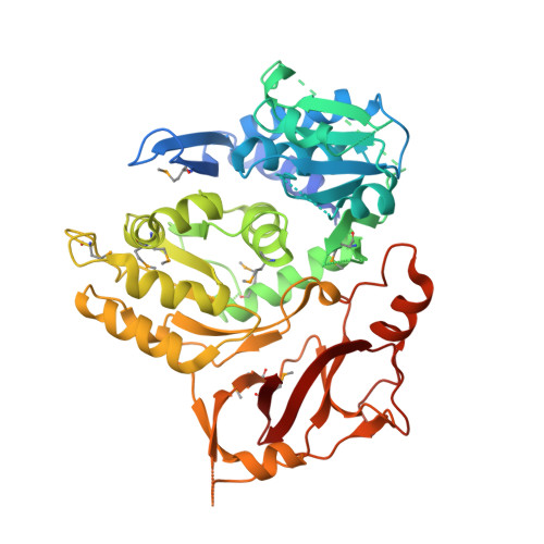
    3E53

  • PubMed Abstract: 

    The recent discovery of fatty acyl-AMP ligases (FAALs) in Mycobacterium tuberculosis (Mtb) provided a new perspective of fatty acid activation. These proteins convert fatty acids to the corresponding adenylates, which are intermediates of acyl-CoA-synthesizing fatty acyl-CoA ligases (FACLs). Presently, it is not evident how obligate pathogens such as Mtb have evolved such new themes of functional versatility and whether the activation of fatty acids to acyladenylates could indeed be a general mechanism. Here, based on elucidation of the first structure of an FAAL protein and by generating loss-of-function and gain-of-function mutants that interconvert FAAL and FACL activities, we demonstrate that an insertion motif dictates formation of acyladenylate. Because FAALs in Mtb are crucial nodes in the biosynthetic network of virulent lipids, inhibitors directed against these proteins provide a unique multipronged approach to simultaneously disrupting several pathways.


  • Organizational Affiliation
    • National Institute of Immunology, Aruna Asaf Ali Marg, New Delhi 110 067, India.

Macromolecule Content 

  • Total Structure Weight: 52.46 kDa 
  • Atom Count: 3,485 
  • Modeled Residue Count: 425 
  • Deposited Residue Count: 480 
  • Unique protein chains: 1

Macromolecules

Find similar proteins by:|  3D Structure
Entity ID: 1
MoleculeChains  Sequence LengthOrganismDetailsImage
FATTY-ACID-CoA LIGASE FADD28480Mycobacterium tuberculosisMutation(s): 0 
Gene Names: fadD28MT3011Rv2941
EC: 6.2.1 (PDB Primary Data), 6.2.1.49 (UniProt)
UniProt
Find proteins for P9WQ59 (Mycobacterium tuberculosis (strain ATCC 25618 / H37Rv))
Explore P9WQ59 
Go to UniProtKB:  P9WQ59
Entity Groups
Sequence Clusters30% Identity50% Identity70% Identity90% Identity95% Identity100% Identity
UniProt GroupP9WQ59
Sequence Annotations
Expand
Reference Sequence

Small Molecules

Modified Residues  1 Unique
IDChains TypeFormula2D DiagramParent
MSE
Query on MSE
A
L-PEPTIDE LINKINGC5 H11 N O2 SeMET

Experimental Data & Validation

Experimental Data

  • Method: X-RAY DIFFRACTION
  • Resolution: 2.35 Å
  • R-Value Free:  0.267 (Depositor), 0.219 (DCC) 
  • R-Value Work:  0.207 (Depositor), 0.210 (DCC) 
  • R-Value Observed: 0.207 (Depositor) 
Space Group: P 21 21 21
Unit Cell:
Length ( Å )Angle ( ˚ )
a = 50.972α = 90
b = 60.74β = 90
c = 136.54γ = 90
Software Package:
Software NamePurpose
CNSrefinement
DENZOdata reduction
SCALEPACKdata scaling
SOLVEphasing

Structure Validation

View Full Validation Report


Entry History 

Deposition Data

Revision History  (Full details and data files)

  • Version 1.0: 2009-01-27
    Type: Initial release
  • Version 1.1: 2011-07-13
    Changes: Version format compliance
  • Version 1.2: 2024-11-20
    Changes: Data collection, Database references, Derived calculations, Structure summary