3E4C | pdb_00003e4c

Procaspase-1 zymogen domain crystal structure


Experimental Data Snapshot

  • Method: X-RAY DIFFRACTION
  • Resolution: 2.05 Å
  • R-Value Free: 
    0.263 (Depositor) 
  • R-Value Work: 
    0.200 (Depositor), 0.212 (DCC) 
  • R-Value Observed: 
    0.204 (Depositor) 

Starting Model: experimental
View more details

wwPDB Validation 3D Report Full Report

Validation slider image for 3E4C

This is version 1.4 of the entry. See complete history

Literature

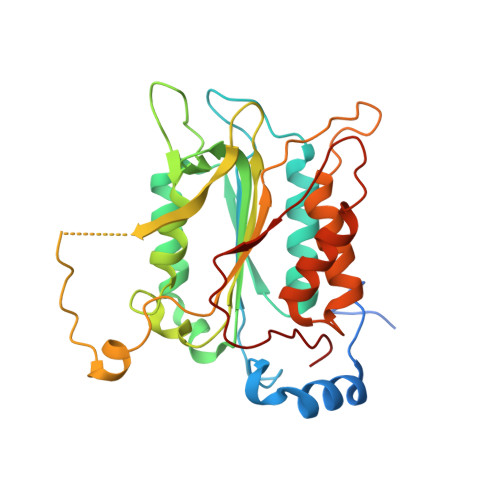
Crystal structure of procaspase-1 zymogen domain reveals insight into inflammatory caspase autoactivation

Elliott, J.M.Rouge, L.Wiesmann, C.Scheer, J.M.

(2009) J Biological Chem 284: 6546-6553

  • DOI: https://doi.org/10.1074/jbc.M806121200
  • Primary Citation Related Structures: 
    3E4C

  • PubMed Abstract: 

    One key event in inflammatory signaling is the activation of the initiator caspase, procaspase-1. Presented here is the crystal structure of the procaspase-1 zymogen without its caspase recruitment domain solved to 2.05 A. Although the isolated domain is monomeric in solution, the protein appeared dimeric in crystals. The loop arrangements in the dimer provide insight into the first autoproteolytic events that occur during activation by oligomerization. Additionally, in contrast to other caspases, we demonstrate that autoproteolysis at the second cleavage site, Asp316, is necessary for conversion to a stable dimer in solution. Critical elements of secondary structure are revealed in the crystal structure that explain why a dimeric protein is favored after proteolysis at this aspartic acid. Dimer stabilization is concurrent with a 130-fold increase in kcat, the sole contributing kinetic factor to an activated and efficient mediator of inflammation.


  • Organizational Affiliation
    • Department of Protein Chemistry, Genentech, Inc., South San Francisco, California 94080, USA.

Macromolecule Content 

  • Total Structure Weight: 67.62 kDa 
  • Atom Count: 4,423 
  • Modeled Residue Count: 540 
  • Deposited Residue Count: 604 
  • Unique protein chains: 1

Macromolecules

Find similar proteins by:|  3D Structure
Entity ID: 1
MoleculeChains  Sequence LengthOrganismDetailsImage
Caspase-1
A, B
302Homo sapiensMutation(s): 1 
Gene Names: CASP1IL1BCIL1BCE
EC: 3.4.22.36
UniProt & NIH Common Fund Data Resources
Find proteins for P29466 (Homo sapiens)
Explore P29466 
Go to UniProtKB:  P29466
PHAROS:  P29466
GTEx:  ENSG00000137752 
Entity Groups
Sequence Clusters30% Identity50% Identity70% Identity90% Identity95% Identity100% Identity
UniProt GroupP29466
Sequence Annotations
Expand
Reference Sequence

Small Molecules

Ligands 1 Unique
IDChains Name / Formula / InChI Key2D Diagram3D Interactions
MG

Query on MG


Download:Ideal Coordinates CCD File
C [auth B]MAGNESIUM ION
Mg
JLVVSXFLKOJNIY-UHFFFAOYSA-N

Experimental Data & Validation

Experimental Data

  • Method: X-RAY DIFFRACTION
  • Resolution: 2.05 Å
  • R-Value Free:  0.263 (Depositor) 
  • R-Value Work:  0.200 (Depositor), 0.212 (DCC) 
  • R-Value Observed: 0.204 (Depositor) 
Space Group: P 1
Unit Cell:
Length ( Å )Angle ( ˚ )
a = 56.1α = 105.95
b = 58.004β = 119.6
c = 60.12γ = 92.77
Software Package:
Software NamePurpose
REFMACrefinement
HKL-2000data collection
HKL-2000data reduction
SCALEPACKdata scaling
PHASERphasing

Structure Validation

View Full Validation Report


Entry History 

Deposition Data

Revision History  (Full details and data files)

  • Version 1.0: 2008-12-30
    Type: Initial release
  • Version 1.1: 2011-07-13
    Changes: Advisory, Refinement description, Version format compliance
  • Version 1.2: 2020-09-09
    Changes: Database references, Derived calculations, Structure summary
  • Version 1.3: 2021-10-20
    Changes: Database references
  • Version 1.4: 2023-08-30
    Changes: Data collection, Refinement description