3E2Y | pdb_00003e2y

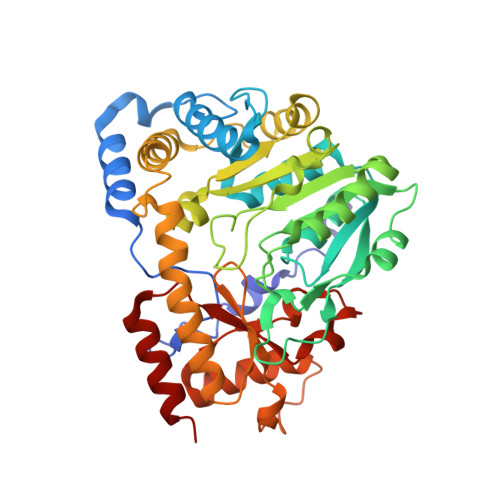
Crystal structure of mouse kynurenine aminotransferase III in complex with glutamine


Experimental Data Snapshot

  • Method: X-RAY DIFFRACTION
  • Resolution: 2.26 Å
  • R-Value Free: 
    0.221 (Depositor), 0.221 (DCC) 
  • R-Value Work: 
    0.175 (Depositor), 0.174 (DCC) 
  • R-Value Observed: 
    0.177 (Depositor) 

Starting Model: experimental
View more details

wwPDB Validation 3D Report Full Report

Validation slider image for 3E2Y

Ligand Structure Quality Assessment 


This is version 1.3 of the entry. See complete history. 

Re-refinement Note

A newer entry is available that reflects an alternative modeling of the original data: 5VEQ

Literature

Correction for Han et al., "Biochemical and Structural Properties of Mouse Kynurenine Aminotransferase III".

Han, Q., Robinson, H., Cai, T., Tagle, D.A., Li, J.

(2018) Mol Cell Biol 38

  • DOI: https://doi.org/10.1128/MCB.00099-18
  • Primary Citation Related Structures: 
    3E2F, 3E2Y, 3E2Z

  • Organizational Affiliation: 
    • Department of Biochemistry, Virginia Tech, Blacksburg, Virginia 24061.

Macromolecule Content 

  • Total Structure Weight: 93.61 kDa 
  • Atom Count: 6,993 
  • Modeled Residue Count: 820 
  • Deposited Residue Count: 820 
  • Unique protein chains: 1

Macromolecules

Find similar proteins by:|  3D Structure
Entity ID: 1
MoleculeChains  Sequence LengthOrganismDetailsImage
Kynurenine-oxoglutarate transaminase 3
A, B
410Mus musculusMutation(s): 0 
Gene Names: Ccbl2, Kat3
EC: 2.6.1.7 (PDB Primary Data), 4.4.1.13 (PDB Primary Data), 2.6.1.63 (UniProt)
UniProt & NIH Common Fund Data Resources
Find proteins for Q71RI9 (Mus musculus)
Explore Q71RI9 
Go to UniProtKB:  Q71RI9
IMPC:  MGI:2677849
Entity Groups
Sequence Clusters30% Identity50% Identity70% Identity90% Identity95% Identity100% Identity
UniProt GroupQ71RI9
Sequence Annotations
Expand
Reference Sequence

Small Molecules

Ligands 3 Unique
IDChains Name / Formula / InChI Key2D Diagram3D Interactions
PMP

Query on PMP


Download:Ideal Coordinates CCD File
D [auth A],
G [auth B]
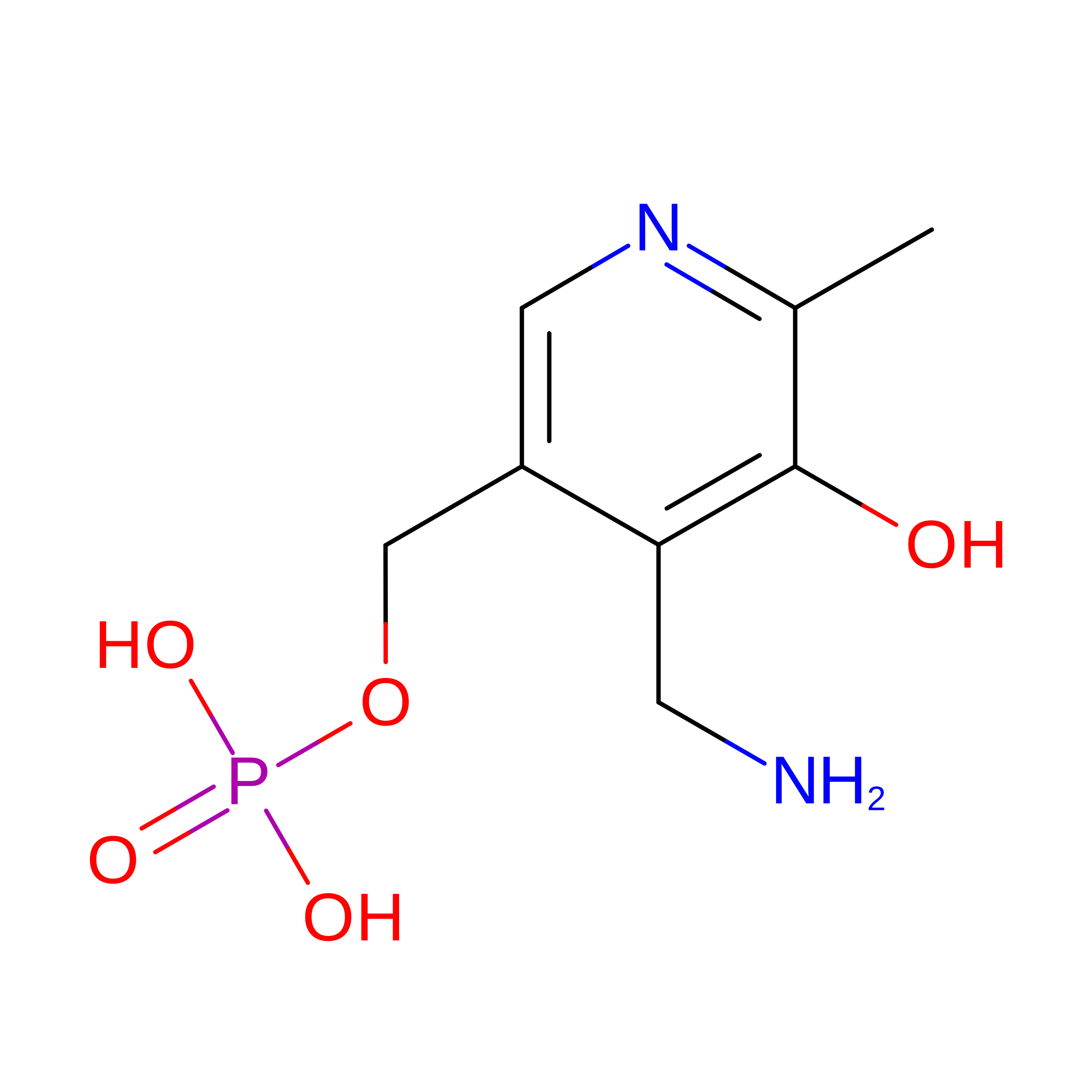
4'-DEOXY-4'-AMINOPYRIDOXAL-5'-PHOSPHATE
C8 H13 N2 O5 P
ZMJGSOSNSPKHNH-UHFFFAOYSA-N
GLN

Query on GLN


Download:Ideal Coordinates CCD File
C [auth A],
H [auth B]
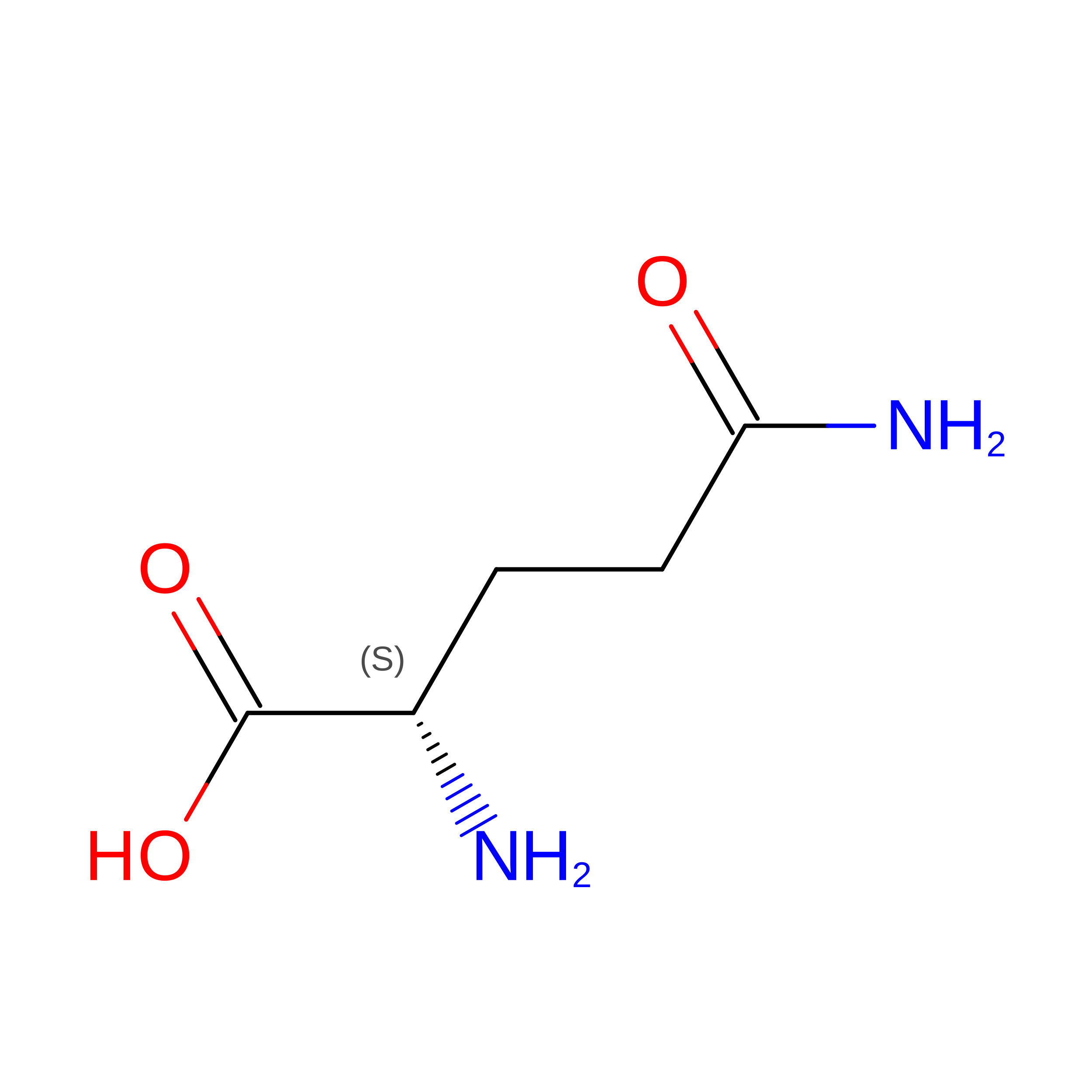
GLUTAMINE
C5 H10 N2 O3
ZDXPYRJPNDTMRX-VKHMYHEASA-N
GOL

Query on GOL


Download:Ideal Coordinates CCD File
E [auth A],
F [auth A],
I [auth B],
J [auth B]
GLYCEROL
C3 H8 O3
PEDCQBHIVMGVHV-UHFFFAOYSA-N

Experimental Data & Validation

Experimental Data

  • Method: X-RAY DIFFRACTION
  • Resolution: 2.26 Å
  • R-Value Free:  0.221 (Depositor), 0.221 (DCC) 
  • R-Value Work:  0.175 (Depositor), 0.174 (DCC) 
  • R-Value Observed: 0.177 (Depositor) 
Space Group: P 43 21 2
Unit Cell:
Length ( Å )Angle ( ˚ )
a = 91.567α = 90
b = 91.567β = 90
c = 232.536γ = 90
Software Package:
Software NamePurpose
REFMACrefinement
HKL-2000data collection
DENZOdata reduction
SCALAdata scaling
MOLREPphasing

Structure Validation

View Full Validation Report


Ligand Structure Quality Assessment 


Entry History 

Deposition Data

Revision History  (Full details and data files)

  • Version 1.0: 2008-12-30
    Type: Initial release
  • Version 1.1: 2011-07-13
    Changes: Non-polymer description, Version format compliance
  • Version 1.2: 2018-08-01
    Changes: Advisory, Data collection, Database references
  • Version 1.3: 2023-08-30
    Changes: Data collection, Database references, Derived calculations, Refinement description