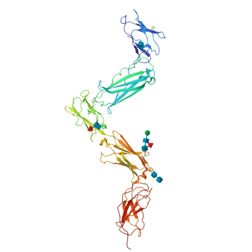
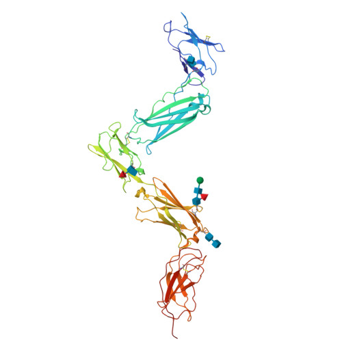
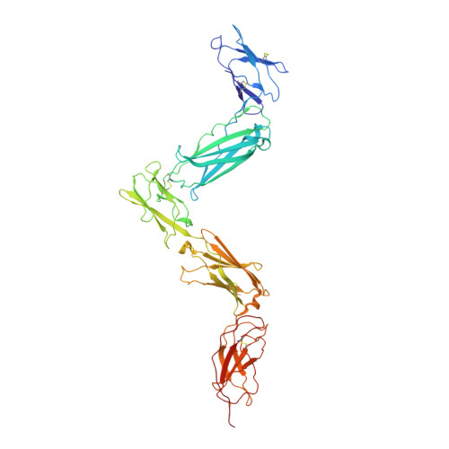
3E0G

Structure of the Leukemia Inhibitory Factor Receptor (LIF-R) domains D1-D5


Experimental Data Snapshot

  • Method: X-RAY DIFFRACTION
  • Resolution: 3.10 Å
  • R-Value Free: 0.309 
  • R-Value Work: 0.252 
  • R-Value Observed: 0.255 

Starting Model: experimental
View more details

wwPDB Validation   3D Report Full Report


This is version 2.2 of the entry. See complete history


Literature

Structural organization of a full-length gp130/LIF-R cytokine receptor transmembrane complex.

Skiniotis, G.Lupardus, P.J.Martick, M.Walz, T.Garcia, K.C.

(2008) Mol Cell 31: 737-748

  • DOI: https://doi.org/10.1016/j.molcel.2008.08.011
  • Primary Citation of Related Structures:  
    3E0G

  • PubMed Abstract: 

    gp130 is a shared receptor for at least nine cytokines and can signal either as a homodimer or as a heterodimer with Leukemia Inhibitory Factor Receptor (LIF-R). Here, we biophysically and structurally characterize the full-length, transmembrane form of a quaternary cytokine receptor complex consisting of gp130, LIF-R, the cytokine Ciliary Neurotrophic Factor (CNTF), and its alpha receptor (CNTF-Ralpha). Thermodynamic analysis indicates that, unlike the cooperative assembly of the symmetric gp130/Interleukin-6/IL-6Ralpha hexameric complex, CNTF/CNTF-Ralpha heterodimerizes gp130 and LIF-R via noncooperative energetics to form an asymmetric 1:1:1:1 complex. Single particle electron microscopic analysis of the full-length gp130/LIF-R/CNTF-Ralpha/CNTF quaternary complex elucidates an asymmetric structural arrangement, in which the receptor extracellular and transmembrane segments join as a continuous, rigid unit, poised to sensitively transduce ligand engagement to the membrane-proximal intracellular signaling regions. These studies also enumerate the organizing principles for assembly of the "tall" class of gp130 family cytokine receptor complexes including LIF, IL-27, IL-12, and others.


  • Organizational Affiliation

    Department of Cell Biology, Harvard Medical School, 240 Longwood Avenue, Boston, MA 02115, USA.


Macromolecules
Find similar proteins by:  (by identity cutoff)  |  3D Structure
Entity ID: 1
MoleculeChains Sequence LengthOrganismDetailsImage
Leukemia inhibitory factor receptor483Homo sapiensMutation(s): 6 
Gene Names: LIFR
UniProt & NIH Common Fund Data Resources
Find proteins for P42702 (Homo sapiens)
Explore P42702 
Go to UniProtKB:  P42702
PHAROS:  P42702
GTEx:  ENSG00000113594 
Entity Groups  
Sequence Clusters30% Identity50% Identity70% Identity90% Identity95% Identity100% Identity
UniProt GroupP42702
Glycosylation
Glycosylation Sites: 4Go to GlyGen: P42702-1
Sequence Annotations
Expand
  • Reference Sequence
Oligosaccharides

Help

Entity ID: 2
MoleculeChains Length2D Diagram Glycosylation3D Interactions
alpha-D-mannopyranose-(1-4)-2-acetamido-2-deoxy-beta-D-glucopyranose-(1-4)-[alpha-L-fucopyranose-(1-6)]2-acetamido-2-deoxy-beta-D-glucopyranose
B
4N-Glycosylation
Glycosylation Resources
GlyTouCan:  G47477HI
GlyCosmos:  G47477HI
GlyGen:  G47477HI
Entity ID: 3
MoleculeChains Length2D Diagram Glycosylation3D Interactions
2-acetamido-2-deoxy-beta-D-glucopyranose-(1-4)-2-acetamido-2-deoxy-beta-D-glucopyranose
C, E
2N-Glycosylation
Glycosylation Resources
GlyTouCan:  G42666HT
GlyCosmos:  G42666HT
GlyGen:  G42666HT
Entity ID: 4
MoleculeChains Length2D Diagram Glycosylation3D Interactions
2-acetamido-2-deoxy-beta-D-glucopyranose-(1-4)-[alpha-L-fucopyranose-(1-6)]2-acetamido-2-deoxy-beta-D-glucopyranose
D
3N-Glycosylation
Glycosylation Resources
GlyTouCan:  G21290RB
GlyCosmos:  G21290RB
GlyGen:  G21290RB
Experimental Data & Validation

Experimental Data

  • Method: X-RAY DIFFRACTION
  • Resolution: 3.10 Å
  • R-Value Free: 0.309 
  • R-Value Work: 0.252 
  • R-Value Observed: 0.255 
  • Space Group: C 1 2 1
Unit Cell:
Length ( Å )Angle ( ˚ )
a = 90.008α = 90
b = 143.169β = 110.38
c = 80.362γ = 90
Software Package:
Software NamePurpose
REFMACrefinement
HKL-2000data collection
MOSFLMdata reduction
SCALAdata scaling
PHASERphasing

Structure Validation

View Full Validation Report


Entry History 

Deposition Data

Revision History  (Full details and data files)

  • Version 1.0: 2008-09-23
    Type: Initial release
  • Version 1.1: 2011-07-13
    Changes: Advisory, Version format compliance
  • Version 2.0: 2020-07-29
    Type: Remediation
    Reason: Carbohydrate remediation
    Changes: Advisory, Atomic model, Data collection, Derived calculations, Structure summary
  • Version 2.1: 2021-10-20
    Changes: Database references, Structure summary
  • Version 2.2: 2024-10-30
    Changes: Data collection, Refinement description, Structure summary