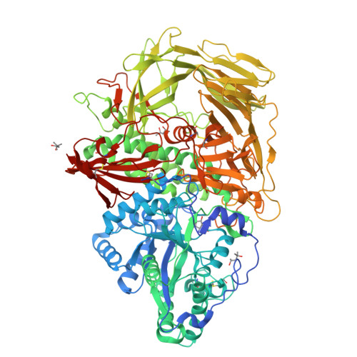
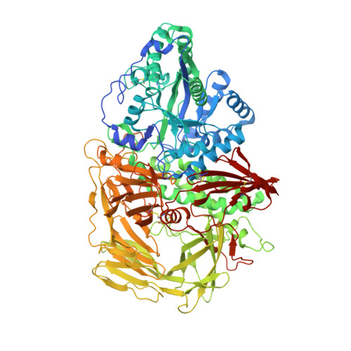
3DX2

Golgi mannosidase II complex with MANNOSTATIN B


Experimental Data Snapshot

  • Method: X-RAY DIFFRACTION
  • Resolution: 1.40 Å
  • R-Value Free: 0.164 
  • R-Value Work: 0.142 
  • R-Value Observed: 0.142 

Starting Model: experimental
View more details

wwPDB Validation   3D Report Full Report


Ligand Structure Quality Assessment 

Created with Raphaël 2.3.0Worse 01 BetterLigand structure goodness of fit to experimental dataBest fitted MZBClick on this verticalbar to view details

This is version 1.4 of the entry. See complete history


Literature

The molecular basis of inhibition of Golgi alpha-mannosidase II by mannostatin A.

Kuntz, D.A.Zhong, W.Guo, J.Rose, D.R.Boons, G.J.

(2009) Chembiochem 10: 268-277

  • DOI: https://doi.org/10.1002/cbic.200800538
  • Primary Citation of Related Structures:  
    3DX0, 3DX1, 3DX2, 3DX3, 3DX4

  • PubMed Abstract: 

    Mannostatin A is a potent inhibitor of the mannose-trimming enzyme, Golgi alpha-mannosidase II (GMII), which acts late in the N-glycan processing pathway. Inhibition of this enzyme provides a route to blocking the transformation-associated changes in cancer cell surface oligosaccharide structures. Here, we report on the synthesis of new Mannostatin derivatives and analyze their binding in the active site of Drosophila GMII by X-ray crystallography. The results indicate that the interaction with the backbone carbonyl of Arg876 is crucial to the high potency of the inhibitor-an effect enhanced by the hydrophobic interaction between the thiomethyl group and an aromatic pocket vicinal to the cleavage site. The various structures indicate that differences in the hydration of protein-ligand complexes are also important determinants of plasticity as well as selectivity of inhibitor binding.


  • Organizational Affiliation

    Ontario Cancer Institute and Department of Medical Biophysics, University of Toronto, Toronto, Ontario M5G 1L7, Canada.


Macromolecules
Find similar proteins by:  (by identity cutoff)  |  3D Structure
Entity ID: 1
MoleculeChains Sequence LengthOrganismDetailsImage
Alpha-mannosidase 21,045Drosophila melanogasterMutation(s): 0 
Gene Names: alpha-Man-IIGmIICG18802
EC: 3.2.1.114
UniProt
Find proteins for Q24451 (Drosophila melanogaster)
Explore Q24451 
Go to UniProtKB:  Q24451
Entity Groups  
Sequence Clusters30% Identity50% Identity70% Identity90% Identity95% Identity100% Identity
UniProt GroupQ24451
Sequence Annotations
Expand
  • Reference Sequence
Small Molecules
Ligands 4 Unique
IDChains Name / Formula / InChI Key2D Diagram3D Interactions
MZB
Query on MZB

Download Ideal Coordinates CCD File 
D [auth A](1R,2R,3R,4S,5R)-4-amino-5-[(R)-methylsulfinyl]cyclopentane-1,2,3-triol
C6 H13 N O4 S
ZJFKRRRXLLAUHQ-DMRLTRFVSA-N
MPD
Query on MPD

Download Ideal Coordinates CCD File 
E [auth A],
F [auth A]
(4S)-2-METHYL-2,4-PENTANEDIOL
C6 H14 O2
SVTBMSDMJJWYQN-YFKPBYRVSA-N
MRD
Query on MRD

Download Ideal Coordinates CCD File 
C [auth A](4R)-2-METHYLPENTANE-2,4-DIOL
C6 H14 O2
SVTBMSDMJJWYQN-RXMQYKEDSA-N
ZN
Query on ZN

Download Ideal Coordinates CCD File 
B [auth A]ZINC ION
Zn
PTFCDOFLOPIGGS-UHFFFAOYSA-N
Binding Affinity Annotations 
IDSourceBinding Affinity
MZB PDBBind:  3DX2 Ki: 150 (nM) from 1 assay(s)
Experimental Data & Validation

Experimental Data

  • Method: X-RAY DIFFRACTION
  • Resolution: 1.40 Å
  • R-Value Free: 0.164 
  • R-Value Work: 0.142 
  • R-Value Observed: 0.142 
  • Space Group: P 21 21 21
Unit Cell:
Length ( Å )Angle ( ˚ )
a = 68.63α = 90
b = 109.281β = 90
c = 137.77γ = 90
Software Package:
Software NamePurpose
REFMACrefinement
CNSrefinement
PDB_EXTRACTdata extraction
ADSCdata collection
HKL-2000data reduction
SADABSdata scaling
CNSphasing

Structure Validation

View Full Validation Report


Ligand Structure Quality Assessment 

Created with Raphaël 2.3.0Worse 01 BetterLigand structure goodness of fit to experimental dataBest fitted MZBClick on this verticalbar to view details

Entry History 

Deposition Data

Revision History  (Full details and data files)

  • Version 1.0: 2009-07-07
    Type: Initial release
  • Version 1.1: 2011-07-13
    Changes: Version format compliance
  • Version 1.2: 2019-07-24
    Changes: Data collection, Refinement description
  • Version 1.3: 2023-08-30
    Changes: Data collection, Database references, Derived calculations, Refinement description
  • Version 1.4: 2024-11-06
    Changes: Structure summary