3DRB | pdb_00003drb

Crystal structure of Human Brain-type Creatine Kinase


Experimental Data Snapshot

  • Method: X-RAY DIFFRACTION
  • Resolution: 2.00 Å
  • R-Value Free: 
    0.227 (Depositor), 0.223 (DCC) 
  • R-Value Work: 
    0.196 (Depositor), 0.192 (DCC) 
  • R-Value Observed: 
    0.212 (Depositor) 

wwPDB Validation 3D Report Full Report

Validation slider image for 3DRB

Ligand Structure Quality Assessment 


This is version 1.3 of the entry. See complete history

Literature

Structural studies of human brain-type creatine kinase complexed with the ADP-Mg2+-NO3- -creatine transition-state analogue complex

Bong, S.M.Moon, J.H.Nam, K.H.Lee, K.S.Chi, Y.M.Hwang, K.Y.

(2008) FEBS Lett 582: 3959-3965

  • DOI: https://doi.org/10.1016/j.febslet.2008.10.039
  • Primary Citation Related Structures: 
    3B6R, 3DRB, 3DRE

  • PubMed Abstract: 

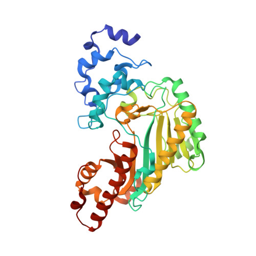
    Creatine kinase is a member of the phosphagen kinase family, which catalyzes the reversible phosphoryl transfer reaction that occurs between ATP and creatine to produce ADP and phosphocreatine. Here, three structural aspects of human-brain-type-creatine-kinase (hBB-CK) were identified by X-ray crystallography: the ligand-free-form at 2.2A; the ADP-Mg2+, nitrate, and creatine complex (transition-state-analogue complex; TSAC); and the ADP-Mg2+-complex at 2.0A. The structures of ligand-bound hBB-CK revealed two different monomeric states in a single homodimer. One monomer is a closed form, either bound to TSAC or the ADP-Mg2+-complex, and the second monomer is an unliganded open form. These structural studies provide a detailed mechanism indicating that the binding of ADP-Mg2+ alone may trigger conformational changes in hBB-CK that were not observed with muscle-type-CK.


  • Organizational Affiliation
    • Division of Biotechnology, College of Life Sciences, Korea University, Seoul 136-713, Republic of Korea.

Macromolecule Content 

  • Total Structure Weight: 85.85 kDa 
  • Atom Count: 6,478 
  • Modeled Residue Count: 752 
  • Deposited Residue Count: 762 
  • Unique protein chains: 1

Macromolecules

Find similar proteins by:|  3D Structure
Entity ID: 1
MoleculeChains  Sequence LengthOrganismDetailsImage
Creatine kinase B-type
A, B
381Homo sapiensMutation(s): 0 
EC: 2.7.3.2
UniProt & NIH Common Fund Data Resources
Find proteins for P12277 (Homo sapiens)
Explore P12277 
Go to UniProtKB:  P12277
PHAROS:  P12277
GTEx:  ENSG00000166165 
Entity Groups
Sequence Clusters30% Identity50% Identity70% Identity90% Identity95% Identity100% Identity
UniProt GroupP12277
Sequence Annotations
Expand
Reference Sequence

Small Molecules

Experimental Data & Validation

Experimental Data

  • Method: X-RAY DIFFRACTION
  • Resolution: 2.00 Å
  • R-Value Free:  0.227 (Depositor), 0.223 (DCC) 
  • R-Value Work:  0.196 (Depositor), 0.192 (DCC) 
  • R-Value Observed: 0.212 (Depositor) 
Space Group: P 43 21 2
Unit Cell:
Length ( Å )Angle ( ˚ )
a = 97.72α = 90
b = 97.72β = 90
c = 166.91γ = 90
Software Package:
Software NamePurpose
DENZOdata reduction
SCALEPACKdata scaling
CNSrefinement
PDB_EXTRACTdata extraction
HKL-2000data collection
HKL-2000data reduction
CNSphasing

Structure Validation

View Full Validation Report


Ligand Structure Quality Assessment 


Entry History 

Deposition Data

Revision History  (Full details and data files)

  • Version 1.0: 2009-03-17
    Type: Initial release
  • Version 1.1: 2011-07-13
    Changes: Version format compliance
  • Version 1.2: 2017-10-25
    Changes: Refinement description
  • Version 1.3: 2024-03-20
    Changes: Data collection, Database references, Derived calculations