3DOO | pdb_00003doo

Crystal structure of shikimate dehydrogenase from Staphylococcus epidermidis complexed with shikimate


Experimental Data Snapshot

  • Method: X-RAY DIFFRACTION
  • Resolution: 2.20 Å
  • R-Value Free: 
    0.264 (Depositor), 0.235 (DCC) 
  • R-Value Work: 
    0.188 (Depositor) 
  • R-Value Observed: 
    0.192 (Depositor) 

Starting Model: experimental
View more details

wwPDB Validation 3D Report Full Report

Validation slider image for 3DOO

Ligand Structure Quality Assessment 


This is version 1.3 of the entry. See complete history

Literature

X-ray crystallographic and enzymatic analyses of shikimate dehydrogenase from Staphylococcus epidermidis

Han, C.Hu, T.Wu, D.Qu, S.Zhou, J.Ding, J.Shen, X.Qu, D.Jiang, H.

(2009) FEBS J 276: 1125-1139

  • DOI: https://doi.org/10.1111/j.1742-4658.2008.06856.x
  • Primary Citation Related Structures: 
    3DON, 3DOO

  • PubMed Abstract: 

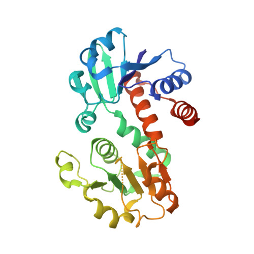
    Shikimate dehydrogenase (SDH) catalyzes the NADPH-dependent reduction of 3-dehydroshikimate to shikimate in the shikimate pathway. In this study, we determined the kinetic properties and crystal structures of Staphylococcus epidermidis SDH (SeSDH) both in its ligand-free form and in complex with shikimate. SeSDH has a k(cat) of 22.8 s(-1) and a K(m) of 73 mum towards shikimate, and a K(m) of 100 microM towards NADP. The overall folding of SeSDH comprises the N-terminal alpha/beta domain for substrate binding and the C-terminal Rossmann fold for NADP binding. The active site is within a large groove between the two domains. Residue Tyr211, normally regarded as important for substrate binding, does not interact with shikimate in the binary SeSDH-shikimate complex structure. However, the Y211F mutation leads to a significant decrease in k(cat) and a minor increase in the K(m) for shikimate. The results indicate that the main function of Tyr211 may be to stabilize the catalytic intermediate during catalysis. The NADP-binding domain of SeSDH is less conserved. The usually long helix specifically recognizing the adenine ribose phosphate is substituted with a short 3(10) helix in the NADP-binding domain. Moreover, the interdomain angle of SeSDH is the widest among all known SDH structures, indicating an inactive 'open' state of the SeSDH structure. Thus, a 'closing' process might occur upon NADP binding to bring the cofactor close to the substrate for catalysis.


  • Organizational Affiliation
    • Institutes of Biomedical Sciences and Key Laboratory of Medical Molecular Virology, Institute of Medical Microbiology, Shanghai Medical College, Fudan University, China.

Macromolecule Content 

  • Total Structure Weight: 31.22 kDa 
  • Atom Count: 2,199 
  • Modeled Residue Count: 258 
  • Deposited Residue Count: 277 
  • Unique protein chains: 1

Macromolecules

Find similar proteins by:|  3D Structure
Entity ID: 1
MoleculeChains  Sequence LengthOrganismDetailsImage
Shikimate dehydrogenase277Staphylococcus epidermidis RP62AMutation(s): 0 
Gene Names: aroE
EC: 1.1.1.25
UniProt
Find proteins for Q5HNV1 (Staphylococcus epidermidis (strain ATCC 35984 / DSM 28319 / BCRC 17069 / CCUG 31568 / BM 3577 / RP62A))
Explore Q5HNV1 
Go to UniProtKB:  Q5HNV1
Entity Groups
Sequence Clusters30% Identity50% Identity70% Identity90% Identity95% Identity100% Identity
UniProt GroupQ5HNV1
Sequence Annotations
Expand
Reference Sequence

Small Molecules

Ligands 1 Unique
IDChains Name / Formula / InChI Key2D Diagram3D Interactions
SKM

Query on SKM


Download:Ideal Coordinates CCD File
B [auth A](3R,4S,5R)-3,4,5-TRIHYDROXYCYCLOHEX-1-ENE-1-CARBOXYLIC ACID
C7 H10 O5
JXOHGGNKMLTUBP-HSUXUTPPSA-N

Experimental Data & Validation

Experimental Data

  • Method: X-RAY DIFFRACTION
  • Resolution: 2.20 Å
  • R-Value Free:  0.264 (Depositor), 0.235 (DCC) 
  • R-Value Work:  0.188 (Depositor) 
  • R-Value Observed: 0.192 (Depositor) 
Space Group: P 1 21 1
Unit Cell:
Length ( Å )Angle ( ˚ )
a = 45.189α = 90
b = 52.529β = 96.14
c = 56.777γ = 90
Software Package:
Software NamePurpose
d*TREKdata scaling
REFMACrefinement
PDB_EXTRACTdata extraction
CNSphasing

Structure Validation

View Full Validation Report


Ligand Structure Quality Assessment 


Entry History 

Deposition Data

Revision History  (Full details and data files)

  • Version 1.0: 2009-05-05
    Type: Initial release
  • Version 1.1: 2011-07-13
    Changes: Version format compliance
  • Version 1.2: 2014-11-12
    Changes: Structure summary
  • Version 1.3: 2023-11-01
    Changes: Data collection, Database references, Derived calculations, Refinement description