3DDL | pdb_00003ddl

Crystallographic Structure of Xanthorhodopsin, a Light-Driven Ion Pump with Dual Chromophore


Experimental Data Snapshot

  • Method: X-RAY DIFFRACTION
  • Resolution: 1.90 Å
  • R-Value Free: 
    0.265 (Depositor), 0.272 (DCC) 
  • R-Value Work: 
    0.247 (Depositor), 0.255 (DCC) 

wwPDB Validation 3D Report Full Report

Validation slider image for 3DDL

Ligand Structure Quality Assessment 


This is version 1.3 of the entry. See complete history

Literature

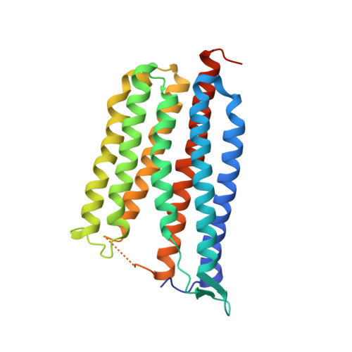
Crystallographic structure of xanthorhodopsin, the light-driven proton pump with a dual chromophore.

Luecke, H.Schobert, B.Stagno, J.Imasheva, E.S.Wang, J.M.Balashov, S.P.Lanyi, J.K.

(2008) Proc Natl Acad Sci U S A 105: 16561-16565

  • DOI: https://doi.org/10.1073/pnas.0807162105
  • Primary Citation Related Structures: 
    3DDL

  • PubMed Abstract: 

    Homologous to bacteriorhodopsin and even more to proteorhodopsin, xanthorhodopsin is a light-driven proton pump that, in addition to retinal, contains a noncovalently bound carotenoid with a function of a light-harvesting antenna. We determined the structure of this eubacterial membrane protein-carotenoid complex by X-ray diffraction, to 1.9-A resolution. Although it contains 7 transmembrane helices like bacteriorhodopsin and archaerhodopsin, the structure of xanthorhodopsin is considerably different from the 2 archaeal proteins. The crystallographic model for this rhodopsin introduces structural motifs for proton transfer during the reaction cycle, particularly for proton release, that are dramatically different from those in other retinal-based transmembrane pumps. Further, it contains a histidine-aspartate complex for regulating the pK(a) of the primary proton acceptor not present in archaeal pumps but apparently conserved in eubacterial pumps. In addition to aiding elucidation of a more general proton transfer mechanism for light-driven energy transducers, the structure defines also the geometry of the carotenoid and the retinal. The close approach of the 2 polyenes at their ring ends explains why the efficiency of the excited-state energy transfer is as high as approximately 45%, and the 46 degrees angle between them suggests that the chromophore location is a compromise between optimal capture of light of all polarization angles and excited-state energy transfer.


  • Organizational Affiliation
    • Departments of Molecular Biology and Biochemistry, and Physiology and Biophysics, and Center for Biomembrane Systems, University of California, Irvine, CA 92697.

Macromolecule Content 

  • Total Structure Weight: 64.18 kDa 
  • Atom Count: 4,417 
  • Modeled Residue Count: 502 
  • Deposited Residue Count: 546 
  • Unique protein chains: 1

Macromolecules

Find similar proteins by:|  3D Structure
Entity ID: 1
MoleculeChains  Sequence LengthOrganismDetailsImage
Xanthorhodopsin
A, B
273Salinibacter ruberMutation(s): 0 
Membrane Entity: Yes 
UniProt
Find proteins for Q2S2F8 (Salinibacter ruber (strain DSM 13855 / M31))
Explore Q2S2F8 
Go to UniProtKB:  Q2S2F8
Entity Groups
Sequence Clusters30% Identity50% Identity70% Identity90% Identity95% Identity100% Identity
UniProt GroupQ2S2F8
Sequence Annotations
Expand
Reference Sequence

Small Molecules

Ligands 5 Unique
IDChains Name / Formula / InChI Key2D Diagram3D Interactions
SXN

Query on SXN


Download:Ideal Coordinates CCD File
C [auth A],
M [auth B]
Salinixanthin
C61 H92 O9
BUNXUZXQWPTVHM-CRAPJSHNSA-N
PCW

Query on PCW


Download:Ideal Coordinates CCD File
CA [auth B]1,2-DIOLEOYL-SN-GLYCERO-3-PHOSPHOCHOLINE
C44 H85 N O8 P
SNKAWJBJQDLSFF-NVKMUCNASA-O
PX4

Query on PX4


Download:Ideal Coordinates CCD File
BA [auth B]1,2-DIMYRISTOYL-SN-GLYCERO-3-PHOSPHOCHOLINE
C36 H73 N O8 P
CITHEXJVPOWHKC-UUWRZZSWSA-O
RET

Query on RET


Download:Ideal Coordinates CCD File
AA [auth B],
L [auth A]
RETINAL
C20 H28 O
NCYCYZXNIZJOKI-OVSJKPMPSA-N
UNL

Query on UNL


Download:Ideal Coordinates CCD File
D [auth A]
E [auth A]
F [auth A]
G [auth A]
H [auth A]
D [auth A],
E [auth A],
F [auth A],
G [auth A],
H [auth A],
I [auth A],
J [auth A],
K [auth A],
N [auth B],
O [auth B],
P [auth B],
Q [auth B],
R [auth B],
S [auth B],
T [auth B],
U [auth B],
V [auth B],
W [auth B],
X [auth B],
Y [auth B],
Z [auth B]
Unknown ligand
NCYCYZXNIZJOKI-OVSJKPMPSA-N

Experimental Data & Validation

Experimental Data

  • Method: X-RAY DIFFRACTION
  • Resolution: 1.90 Å
  • R-Value Free:  0.265 (Depositor), 0.272 (DCC) 
  • R-Value Work:  0.247 (Depositor), 0.255 (DCC) 
Space Group: P 1
Unit Cell:
Length ( Å )Angle ( ˚ )
a = 52.74α = 76.35
b = 59.49β = 74.93
c = 59.72γ = 64.08
Software Package:
Software NamePurpose
d*TREKdata scaling
CNSrefinement
PDB_EXTRACTdata extraction
Blu-Icedata collection
d*TREKdata reduction
PHASERphasing

Structure Validation

View Full Validation Report


Ligand Structure Quality Assessment 


Entry History 

Deposition Data

Revision History  (Full details and data files)

  • Version 1.0: 2008-10-14
    Type: Initial release
  • Version 1.1: 2011-07-13
    Changes: Version format compliance
  • Version 1.2: 2016-06-08
    Changes: Atomic model
  • Version 1.3: 2024-10-30
    Changes: Data collection, Database references, Derived calculations, Structure summary