3DD9 | pdb_00003dd9

Structure of DocH66Y dimer


Experimental Data Snapshot

  • Method: X-RAY DIFFRACTION
  • Resolution: 2.45 Å
  • R-Value Free: 
    0.245 (Depositor), 0.239 (DCC) 
  • R-Value Work: 
    0.211 (Depositor), 0.208 (DCC) 
  • R-Value Observed: 
    0.213 (Depositor) 

Starting Model: experimental
View more details

wwPDB Validation 3D Report Full Report

Validation slider image for 3DD9

This is version 1.4 of the entry. See complete history

Literature

The intrinsically disordered domain of the antitoxin Phd chaperones the toxin Doc against irreversible inactivation and misfolding

De Gieter, S.Konijnenberg, A.Talavera, A.Butterer, A.Haesaerts, S.De Greve, H.Sobott, F.Loris, R.Garcia-Pino, A.

(2014) J Biological Chem 289: 34013-34023

  • DOI: https://doi.org/10.1074/jbc.M114.572396
  • Primary Citation Related Structures: 
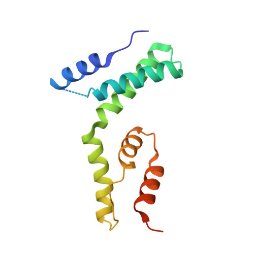
    3DD9

  • PubMed Abstract: 

    The toxin Doc from the phd/doc toxin-antitoxin module targets the cellular translation machinery and is inhibited by its antitoxin partner Phd. Here we show that Phd also functions as a chaperone, keeping Doc in an active, correctly folded conformation. In the absence of Phd, Doc exists in a relatively expanded state that is prone to dimerization through domain swapping with its active site loop acting as hinge region. The domain-swapped dimer is not capable of arresting protein synthesis in vitro, whereas the Doc monomer is. Upon binding to Phd, Doc becomes more compact and is secured in its monomeric state with a neutralized active site.


  • Organizational Affiliation
    • From Structural Biology Brussels, Department of Biotechnology (DBIT), Vrije Universiteit Brussel, Pleinlaan 2, B-1050 Brussels, Belgium, Molecular Recognition Unit (MoRe).

Macromolecule Content 

  • Total Structure Weight: 118.26 kDa 
  • Atom Count: 6,855 
  • Modeled Residue Count: 931 
  • Deposited Residue Count: 1,080 
  • Unique protein chains: 1

Macromolecules

Find similar proteins by:|  3D Structure
Entity ID: 1
MoleculeChains  Sequence LengthOrganismDetailsImage
Death on curing protein
A, B, C, D, E
A, B, C, D, E, F, G, H
135Punavirus P1Mutation(s): 2 
Gene Names: doc
EC: 2.7.11.1
UniProt
Find proteins for Q06259 (Escherichia phage P1)
Explore Q06259 
Go to UniProtKB:  Q06259
Entity Groups
Sequence Clusters30% Identity50% Identity70% Identity90% Identity95% Identity100% Identity
UniProt GroupQ06259
Sequence Annotations
Expand
Reference Sequence

Experimental Data & Validation

Experimental Data

  • Method: X-RAY DIFFRACTION
  • Resolution: 2.45 Å
  • R-Value Free:  0.245 (Depositor), 0.239 (DCC) 
  • R-Value Work:  0.211 (Depositor), 0.208 (DCC) 
  • R-Value Observed: 0.213 (Depositor) 
Space Group: P 1 21 1
Unit Cell:
Length ( Å )Angle ( ˚ )
a = 53.084α = 90
b = 197.988β = 93.04
c = 54.109γ = 90
Software Package:
Software NamePurpose
PHASERphasing
PHENIXrefinement
DENZOdata reduction
SCALEPACKdata scaling

Structure Validation

View Full Validation Report


Entry History 

Deposition Data

Revision History  (Full details and data files)

  • Version 1.0: 2009-06-09
    Type: Initial release
  • Version 1.1: 2011-07-13
    Changes: Version format compliance
  • Version 1.2: 2014-07-23
    Changes: Other
  • Version 1.3: 2018-03-07
    Changes: Database references
  • Version 1.4: 2023-11-01
    Changes: Data collection, Database references, Refinement description