3D9B

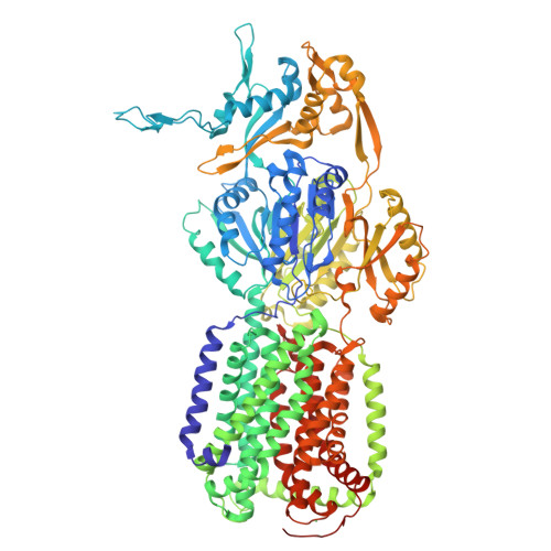
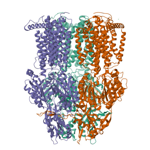
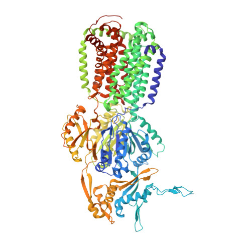
Symmetric structure of E. coli AcrB


Experimental Data Snapshot

  • Method: X-RAY DIFFRACTION
  • Resolution: 3.42 Å
  • R-Value Free: 0.355 
  • R-Value Work: 0.289 
  • R-Value Observed: 0.291 

Starting Model: experimental
View more details

wwPDB Validation   3D Report Full Report


This is version 1.3 of the entry. See complete history


Literature

There is a baby in the bath water: AcrB contamination is a major problem in membrane-protein crystallization.

Veesler, D.Blangy, S.Cambillau, C.Sciara, G.

(2008) Acta Crystallogr Sect F Struct Biol Cryst Commun 64: 880-885

  • DOI: https://doi.org/10.1107/S1744309108028248
  • Primary Citation of Related Structures:  
    3D9B

  • PubMed Abstract: 

    In the course of a crystallographic study of the Methanosarcina mazei CorA transporter, the membrane protein was obtained with at least 95% purity and was submitted to crystallization trials. Small crystals (<100 microm) were grown that diffracted to 3.42 A resolution and belonged to space group R32, with unit-cell parameters a = b = 145.74, c = 514.0 A. After molecular-replacement attempts using available CorA structures as search models failed to yield a solution, it was discovered that the crystals consisted of an Escherichia coli contaminating protein, acriflavine resistance protein B (AcrB), that was present at less than 5% in the protein preparations. AcrB contamination is a major problem when expressing membrane proteins in E. coli since it binds naturally to immobilized metal-ion affinity chromatography (IMAC) resins. Here, the structure is compared with previously deposited AcrB structures and strategies are proposed to avoid this contamination.


  • Organizational Affiliation

    Architecture et Fonction des Macromolécules Biologiques, CNRS et Universités d'Aix-Marseille I et II, UMR 6098, Case 932, 163 Avenue de Luminy, 13288 Marseille CEDEX 9, France. david.veesler@afmb.univ-mrs.fr


Macromolecules
Find similar proteins by:  (by identity cutoff)  |  3D Structure
Entity ID: 1
MoleculeChains Sequence LengthOrganismDetailsImage
Acriflavine resistance protein B1,049Escherichia coli BL21(DE3)Mutation(s): 0 
Membrane Entity: Yes 
UniProt
Find proteins for P31224 (Escherichia coli (strain K12))
Explore P31224 
Go to UniProtKB:  P31224
Entity Groups  
Sequence Clusters30% Identity50% Identity70% Identity90% Identity95% Identity100% Identity
UniProt GroupP31224
Sequence Annotations
Expand
  • Reference Sequence
Small Molecules
Ligands 1 Unique
IDChains Name / Formula / InChI Key2D Diagram3D Interactions
NI
Query on NI

Download Ideal Coordinates CCD File 
B [auth A]NICKEL (II) ION
Ni
VEQPNABPJHWNSG-UHFFFAOYSA-N
Experimental Data & Validation

Experimental Data

  • Method: X-RAY DIFFRACTION
  • Resolution: 3.42 Å
  • R-Value Free: 0.355 
  • R-Value Work: 0.289 
  • R-Value Observed: 0.291 
  • Space Group: H 3 2
Unit Cell:
Length ( Å )Angle ( ˚ )
a = 145.74α = 90
b = 145.74β = 90
c = 514γ = 120
Software Package:
Software NamePurpose
REFMACrefinement
ADSCdata collection
XDSdata reduction
XSCALEdata scaling
MOLREPphasing

Structure Validation

View Full Validation Report


Entry History 

Deposition Data

Revision History  (Full details and data files)

  • Version 1.0: 2008-07-01
    Type: Initial release
  • Version 1.1: 2011-07-13
    Changes: Advisory, Source and taxonomy, Version format compliance
  • Version 1.2: 2012-03-21
    Changes: Database references
  • Version 1.3: 2023-08-30
    Changes: Data collection, Database references, Derived calculations, Refinement description