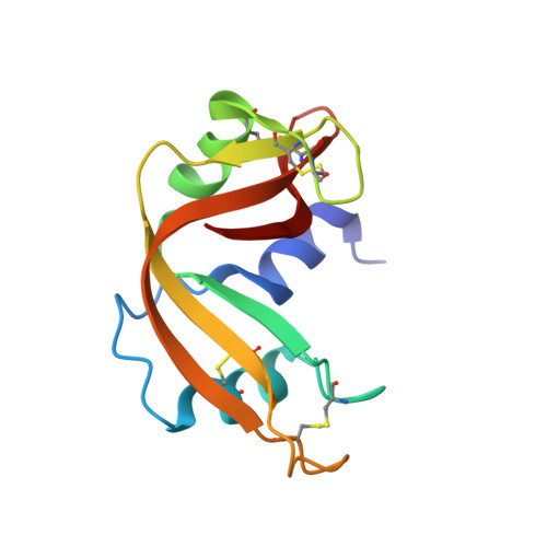
3D8Y

RNase A- 5'-Deoxy-5'-N-piperidinothymidine complex


Experimental Data Snapshot

  • Method: X-RAY DIFFRACTION
  • Resolution: 1.72 Å
  • R-Value Free: 0.230 
  • R-Value Work: 0.189 
  • R-Value Observed: 0.191 

Starting Model: experimental
View more details

wwPDB Validation   3D Report Full Report


Ligand Structure Quality Assessment 


This is version 1.3 of the entry. See complete history


Literature

Morpholino, piperidino, and pyrrolidino derivatives of pyrimidine nucleosides as inhibitors of ribonuclease A: synthesis, biochemical, and crystallographic evaluation

Samanta, A.Leonidas, D.D.Dasgupta, S.Pathak, T.Zographos, S.E.Oikonomakos, N.G.

(2009) J Med Chem 52: 932-942

  • DOI: https://doi.org/10.1021/jm800724t
  • Primary Citation of Related Structures:  
    3D6O, 3D6P, 3D6Q, 3D7B, 3D8Y, 3D8Z

  • PubMed Abstract: 

    Six 5'-deoxy-5'-morpholine, piperidine, and pyrrolidine of pyrimidine nucleosides have been synthesized and characterized. Their inhibitory action to ribonuclease A has been studied by biochemical analysis and X-ray crystallography. These compounds are moderate inhibitors of RNase A with mid-to-upper micromolar inhibition constants (K(i)). The high resolution X-ray crystal structures of the RNase A-inhibitor complexes have shown that all inhibitors bind at the enzyme catalytic cleft with the pyrimidine nucleobase at the B(1)R(2) subsites while the 5' group binds away from the main subsite P(1), where P-O(5') bond cleavage occurs, toward the solvent close to subsite P(0). Structure-activity relationship analysis has demonstrated that the compounds with the larger group in the 5' position are more potent. Comparative structural analysis of these RNase A complexes with other similar RNase A-ligand complexes provides a structural explanation of their potency and suggests ways to improve their efficiency and selectivity. These inhibitors can be the starting point for the development of compounds that can be used as pharmaceuticals against pathologies associated with RNase A homologues such as human angiogenin, which is implicated in tumor induced neovascularization.


  • Organizational Affiliation

    Department of Chemistry, Indian Institute of Technology, Kharagpur 721302, India.


Macromolecules
Find similar proteins by:  (by identity cutoff)  |  3D Structure
Entity ID: 1
MoleculeChains Sequence LengthOrganismDetailsImage
Ribonuclease pancreatic
A, B
124Bos taurusMutation(s): 0 
EC: 3.1.27.5 (PDB Primary Data), 4.6.1.18 (UniProt)
UniProt
Find proteins for P61823 (Bos taurus)
Explore P61823 
Go to UniProtKB:  P61823
Entity Groups  
Sequence Clusters30% Identity50% Identity70% Identity90% Identity95% Identity100% Identity
UniProt GroupP61823
Sequence Annotations
Expand
  • Reference Sequence
Small Molecules
Binding Affinity Annotations 
IDSourceBinding Affinity
T3S BindingDB:  3D8Y Ki: 3.96e+5 (nM) from 1 assay(s)
PDBBind:  3D8Y Ki: 3.96e+5 (nM) from 1 assay(s)
Experimental Data & Validation

Experimental Data

  • Method: X-RAY DIFFRACTION
  • Resolution: 1.72 Å
  • R-Value Free: 0.230 
  • R-Value Work: 0.189 
  • R-Value Observed: 0.191 
  • Space Group: C 1 2 1
Unit Cell:
Length ( Å )Angle ( ˚ )
a = 100.161α = 90
b = 32.421β = 90.9
c = 72.567γ = 90
Software Package:
Software NamePurpose
REFMACrefinement
ADSCdata collection
DENZOdata reduction
SCALEPACKdata scaling
REFMACphasing

Structure Validation

View Full Validation Report


Ligand Structure Quality Assessment 


Entry History 

Deposition Data

Revision History  (Full details and data files)

  • Version 1.0: 2009-02-10
    Type: Initial release
  • Version 1.1: 2011-07-13
    Changes: Advisory, Version format compliance
  • Version 1.2: 2014-01-29
    Changes: Database references
  • Version 1.3: 2023-11-01
    Changes: Data collection, Database references, Derived calculations, Refinement description