3D7O | pdb_00003d7o

Human hemoglobin, nitrogen dioxide anion modified


Experimental Data Snapshot

  • Method: X-RAY DIFFRACTION
  • Resolution: 1.80 Å
  • R-Value Free: 
    0.237 (Depositor), 0.238 (DCC) 
  • R-Value Work: 
    0.206 (Depositor), 0.207 (DCC) 
  • R-Value Observed: 
    0.208 (Depositor) 

Starting Model: experimental
View more details

wwPDB Validation 3D Report Full Report

Validation slider image for 3D7O

Ligand Structure Quality Assessment 


This is version 1.2 of the entry. See complete history

Literature

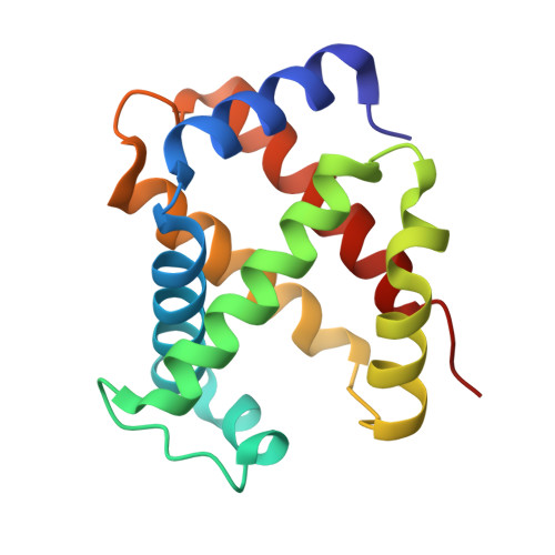
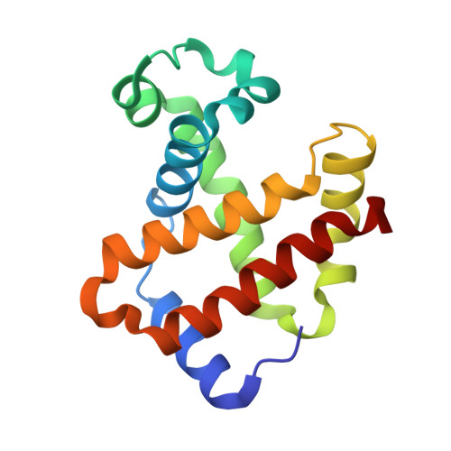
The nitrite anion binds to human hemoglobin via the uncommon O-nitrito mode.

Yi, J.Safo, M.K.Richter-Addo, G.B.

(2008) Biochemistry 47: 8247-8249

  • DOI: https://doi.org/10.1021/bi801015c
  • Primary Citation Related Structures: 
    3D7O

  • PubMed Abstract: 

    The nitrite anion is known to oxidize and degrade hemoglobin (Hb). Recent literature reports suggest a nitrite reductase activity for Hb, converting nitrite into nitric oxide. Surprisingly, no structural information about Hb-nitrite interactions has been reported. We have determined the crystal structure of the ferric Hb-nitrite complex at 1.80 A resolution. The nitrite ligand adopts the uncommon O-nitrito binding mode. In addition, the nitrito conformations in the alpha and beta subunits are different, reflecting subtle effects of the distal His in orienting the nitrite ligand in the O-nitrito binding mode.


  • Organizational Affiliation
    • Department of Chemistry and Biochemistry, University of Oklahoma, Norman, Oklahoma 73019, USA.

Macromolecule Content 

  • Total Structure Weight: 32.46 kDa 
  • Atom Count: 2,468 
  • Modeled Residue Count: 287 
  • Deposited Residue Count: 287 
  • Unique protein chains: 2

Macromolecules

Find similar proteins by:|  3D Structure
Entity ID: 1
MoleculeChains  Sequence LengthOrganismDetailsImage
Hemoglobin subunit alpha141Homo sapiensMutation(s): 0 
UniProt & NIH Common Fund Data Resources
Find proteins for P69905 (Homo sapiens)
Explore P69905 
Go to UniProtKB:  P69905
Entity Groups
Sequence Clusters30% Identity50% Identity70% Identity90% Identity95% Identity100% Identity
UniProt GroupP69905
Sequence Annotations
Expand
Reference Sequence
Find similar proteins by:|  3D Structure
Entity ID: 2
MoleculeChains  Sequence LengthOrganismDetailsImage
Hemoglobin subunit beta146Homo sapiensMutation(s): 0 
UniProt & NIH Common Fund Data Resources
Find proteins for P68871 (Homo sapiens)
Explore P68871 
Go to UniProtKB:  P68871
PHAROS:  P68871
GTEx:  ENSG00000244734 
Entity Groups
Sequence Clusters30% Identity50% Identity70% Identity90% Identity95% Identity100% Identity
UniProt GroupP68871
Sequence Annotations
Expand
Reference Sequence

Experimental Data & Validation

Experimental Data

  • Method: X-RAY DIFFRACTION
  • Resolution: 1.80 Å
  • R-Value Free:  0.237 (Depositor), 0.238 (DCC) 
  • R-Value Work:  0.206 (Depositor), 0.207 (DCC) 
  • R-Value Observed: 0.208 (Depositor) 
Space Group: P 41 21 2
Unit Cell:
Length ( Å )Angle ( ˚ )
a = 53.538α = 90
b = 53.538β = 90
c = 191.77γ = 90
Software Package:
Software NamePurpose
d*TREKdata scaling
MOLREPphasing
REFMACrefinement
PDB_EXTRACTdata extraction
CrystalCleardata collection
d*TREKdata reduction

Structure Validation

View Full Validation Report


Ligand Structure Quality Assessment 


Entry History 

Deposition Data

Revision History  (Full details and data files)

  • Version 1.0: 2009-05-05
    Type: Initial release
  • Version 1.1: 2011-07-13
    Changes: Version format compliance
  • Version 1.2: 2023-08-30
    Changes: Data collection, Database references, Derived calculations, Refinement description