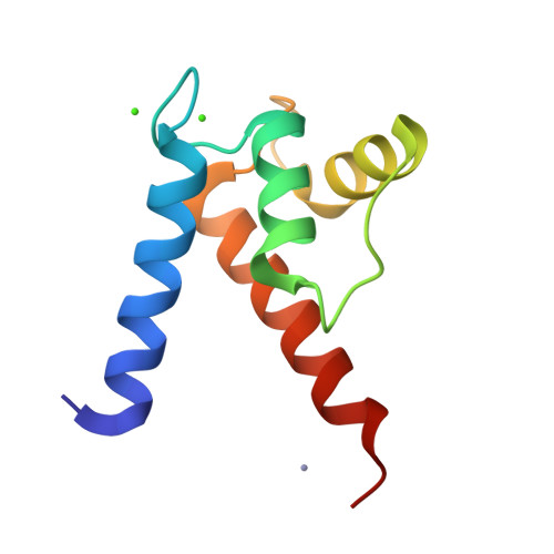
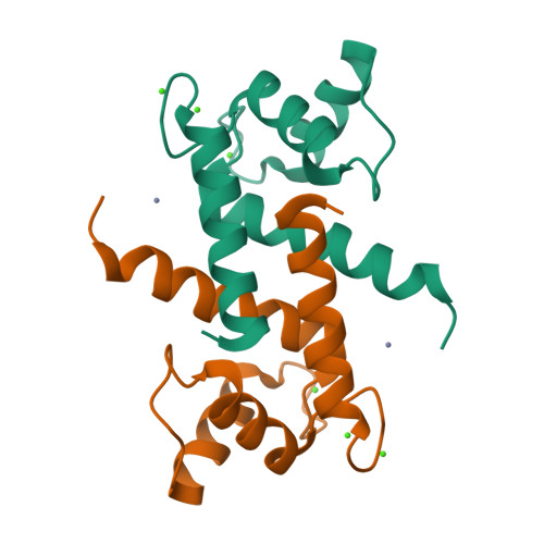
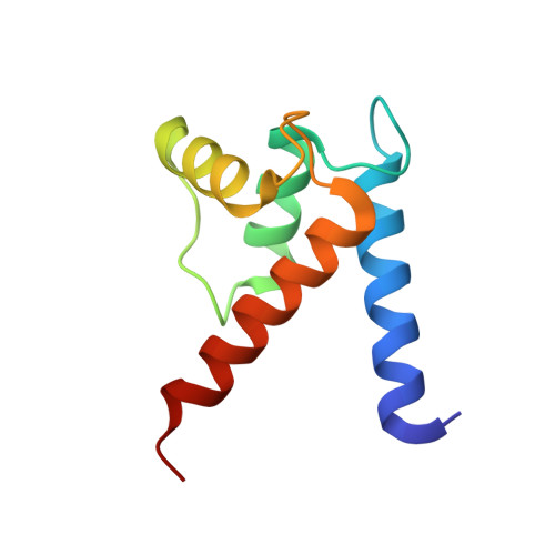
The crystal structures of human S100B in the zinc- and calcium-loaded state at three pH values reveal zinc ligand swapping.
Ostendorp, T., Diez, J., Heizmann, C.W., Fritz, G.(2011) Biochim Biophys Acta 1813: 1083-1091
- PubMed: 20950652
- DOI: https://doi.org/10.1016/j.bbamcr.2010.10.006
- Primary Citation of Related Structures:
3CZT, 3D0Y, 3D10 - PubMed Abstract:
S100B is a homodimeric zinc-, copper-, and calcium-binding protein of the family of EF-hand S100 proteins. Zn(2+) binding to S100B increases its affinity towards Ca(2+) as well as towards target peptides and proteins. Cu(2+) and Zn(2+) bind presumably to the same site in S100B. We determined the structures of human Zn(2+)- and Ca(2+)-loaded S100B at pH 6.5, pH 9, and pH 10 by X-ray crystallography at 1.5, 1.4, and 1.65Å resolution, respectively. Two Zn(2+) ions are coordinated tetrahedrally at the dimer interface by His and Glu residues from both subunits. The crystal structures revealed that ligand swapping occurs for one of the four ligands in the Zn(2+)-binding sites. Whereas at pH 9, the Zn(2+) ions are coordinated by His15, His25, His 85', and His 90', at pH 6.5 and pH 10, His90' is replaced by Glu89'. The results document that the Zn(2+)-binding sites are flexible to accommodate other metal ions such as Cu(2+). Moreover, we characterized the structural changes upon Zn(2+) binding, which might lead to increased affinity towards Ca(2+) as well as towards target proteins. We observed that in Zn(2+)-Ca(2+)-loaded S100B the C-termini of helix IV adopt a distinct conformation. Zn(2+) binding induces a repositioning of residues Phe87 and Phe88, which are involved in target protein binding. This article is part of a Special Issue entitled: 11th European Symposium on Calcium.
Organizational Affiliation:
Fachbereich Biologie, Mathematisch-Naturwissenschaftliche Sektion, Universität Konstanz, 78459 Konstanz, Germany.


