3CZ9 | pdb_00003cz9

Crystal Structure of E18L DJ-1


Experimental Data Snapshot

  • Method: X-RAY DIFFRACTION
  • Resolution: 1.15 Å
  • R-Value Free: 
    0.140 (Depositor), 0.128 (DCC) 
  • R-Value Work: 
    0.115 (Depositor), 0.122 (DCC) 
  • R-Value Observed: 
    0.115 (Depositor) 

Starting Model: experimental
View more details

wwPDB Validation 3D Report Full Report

Validation slider image for 3CZ9

Ligand Structure Quality Assessment 


This is version 1.3 of the entry. See complete history

Literature

Cysteine pKa depression by a protonated glutamic acid in human DJ-1.

Witt, A.C.Lakshminarasimhan, M.Remington, B.C.Hasim, S.Pozharski, E.Wilson, M.A.

(2008) Biochemistry 47: 7430-7440

  • DOI: https://doi.org/10.1021/bi800282d
  • Primary Citation Related Structures: 
    2OR3, 3CY6, 3CYF, 3CZ9, 3CZA

  • PubMed Abstract: 

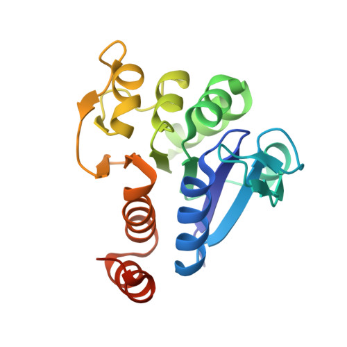
    Human DJ-1, a disease-associated protein that protects cells from oxidative stress, contains an oxidation-sensitive cysteine (C106) that is essential for its cytoprotective activity. The origin of C106 reactivity is obscure, due in part to the absence of an experimentally determined p K a value for this residue. We have used atomic-resolution X-ray crystallography and UV spectroscopy to show that C106 has a depressed p K a of 5.4 +/- 0.1 and that the C106 thiolate accepts a hydrogen bond from a protonated glutamic acid side chain (E18). X-ray crystal structures and cysteine p K a analysis of several site-directed substitutions at residue 18 demonstrate that the protonated carboxylic acid side chain of E18 is required for the maximal stabilization of the C106 thiolate. A nearby arginine residue (R48) participates in a guanidinium stacking interaction with R28 from the other monomer in the DJ-1 dimer and elevates the p K a of C106 by binding an anion that electrostatically suppresses thiol ionization. Our results show that the ionizable residues (E18, R48, and R28) surrounding C106 affect its p K a in a way that is contrary to expectations based on the typical ionization behavior of glutamic acid and arginine. Lastly, a search of the Protein Data Bank (PDB) produces several candidate hydrogen-bonded aspartic/glutamic acid-cysteine interactions, which we propose are particularly common in the DJ-1 superfamily.


  • Organizational Affiliation
    • Department of Biochemistry and Redox Biology Center, The University of Nebraska, Lincoln, Nebraska 68588-0664, USA.

Macromolecule Content 

  • Total Structure Weight: 21.3 kDa 
  • Atom Count: 1,751 
  • Modeled Residue Count: 187 
  • Deposited Residue Count: 197 
  • Unique protein chains: 1

Macromolecules

Find similar proteins by:|  3D Structure
Entity ID: 1
MoleculeChains  Sequence LengthOrganismDetailsImage
Protein DJ-1197Homo sapiensMutation(s): 1 
Gene Names: PARK7
EC: 3.5.1 (UniProt), 3.1.2 (UniProt), 3.5.1.124 (UniProt)
UniProt & NIH Common Fund Data Resources
Find proteins for Q99497 (Homo sapiens)
Explore Q99497 
Go to UniProtKB:  Q99497
PHAROS:  Q99497
GTEx:  ENSG00000116288 
Entity Groups
Sequence Clusters30% Identity50% Identity70% Identity90% Identity95% Identity100% Identity
UniProt GroupQ99497
Sequence Annotations
Expand
Reference Sequence

Small Molecules

Ligands 1 Unique
IDChains Name / Formula / InChI Key2D Diagram3D Interactions
P4C

Query on P4C


Download:Ideal Coordinates CCD File
B [auth A]O-ACETALDEHYDYL-HEXAETHYLENE GLYCOL
C14 H28 O8
CTLLATPOKUEFSQ-UHFFFAOYSA-N

Experimental Data & Validation

Experimental Data

  • Method: X-RAY DIFFRACTION
  • Resolution: 1.15 Å
  • R-Value Free:  0.140 (Depositor), 0.128 (DCC) 
  • R-Value Work:  0.115 (Depositor), 0.122 (DCC) 
  • R-Value Observed: 0.115 (Depositor) 
Space Group: P 31 2 1
Unit Cell:
Length ( Å )Angle ( ˚ )
a = 74.695α = 90
b = 74.695β = 90
c = 74.877γ = 120
Software Package:
Software NamePurpose
SHELXmodel building
SHELXL-97refinement
ADSCdata collection
HKL-2000data reduction
HKL-2000data scaling
PHASERphasing

Structure Validation

View Full Validation Report


Ligand Structure Quality Assessment 


Entry History 

Deposition Data

Revision History  (Full details and data files)

  • Version 1.0: 2008-07-01
    Type: Initial release
  • Version 1.1: 2011-07-13
    Changes: Version format compliance
  • Version 1.2: 2021-10-20
    Changes: Database references, Derived calculations
  • Version 1.3: 2023-08-30
    Changes: Data collection, Refinement description