3CZ4 | pdb_00003cz4

Native AphA class B acid phosphatase/phosphotransferase from E. coli


Experimental Data Snapshot

  • Method: X-RAY DIFFRACTION
  • Resolution: 1.70 Å
  • R-Value Free: 
    0.212 (Depositor), 0.211 (DCC) 
  • R-Value Work: 
    0.181 (Depositor), 0.181 (DCC) 
  • R-Value Observed: 
    0.181 (Depositor) 

Starting Model: experimental
View more details

wwPDB Validation 3D Report Full Report

Validation slider image for 3CZ4

This is version 1.2 of the entry. See complete history

Literature

Structural insights into the catalytic mechanism of the bacterial class B phosphatase AphA belonging to the DDDD superfamily of phosphohydrolases.

Leone, R.Cappelletti, E.Benvenuti, M.Lentini, G.Thaller, M.C.Mangani, S.

(2008) J Mol Biology 384: 478-488

  • DOI: https://doi.org/10.1016/j.jmb.2008.09.050
  • Primary Citation Related Structures: 
    3CZ4

  • PubMed Abstract: 

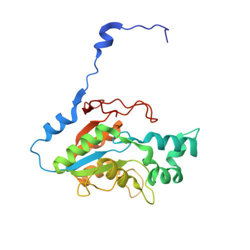
    AphA is a magnesium-dependent, bacterial class B acid phosphatase that catalyzes the hydrolysis of a variety of phosphoester substrates and belongs to the DDDD superfamily of phosphohydrolases. The recently reported crystal structure of AphA from Escherichia coli has revealed the quaternary structure of the enzyme together with hints about its catalytic mechanism. The present work reports the crystal structures of AphA from E. coli in complex with substrate, transition-state, and intermediate analogues. The structures provide new insights into the mechanism of the enzyme and allow a revision of some aspects of the previously proposed mechanism that have broader implications for all the phosphatases of the DDDD superfamily.


  • Organizational Affiliation
    • Dipartimento di Chimica, Università di Siena, Via Aldo Moro 2, I-53100 Siena, Italy.

Macromolecule Content 

  • Total Structure Weight: 24.01 kDa 
  • Atom Count: 2,031 
  • Modeled Residue Count: 209 
  • Deposited Residue Count: 212 
  • Unique protein chains: 1

Macromolecules

Find similar proteins by:|  3D Structure
Entity ID: 1
MoleculeChains  Sequence LengthOrganismDetailsImage
Class B acid phosphatase212Escherichia coli K-12Mutation(s): 0 
Gene Names: aphAnapAyjbPb4055JW4015
EC: 3.1.3.2
UniProt
Find proteins for P0AE22 (Escherichia coli (strain K12))
Explore P0AE22 
Go to UniProtKB:  P0AE22
Entity Groups
Sequence Clusters30% Identity50% Identity70% Identity90% Identity95% Identity100% Identity
UniProt GroupP0AE22
Sequence Annotations
Expand
Reference Sequence

Small Molecules

Ligands 4 Unique
IDChains Name / Formula / InChI Key2D Diagram3D Interactions
EDO

Query on EDO


Download:Ideal Coordinates CCD File
F [auth A],
G [auth A],
H [auth A],
I [auth A],
J [auth A]
1,2-ETHANEDIOL
C2 H6 O2
LYCAIKOWRPUZTN-UHFFFAOYSA-N
ACT

Query on ACT


Download:Ideal Coordinates CCD File
C [auth A]ACETATE ION
C2 H3 O2
QTBSBXVTEAMEQO-UHFFFAOYSA-M
CL

Query on CL


Download:Ideal Coordinates CCD File
D [auth A]CHLORIDE ION
Cl
VEXZGXHMUGYJMC-UHFFFAOYSA-M
MG

Query on MG


Download:Ideal Coordinates CCD File
B [auth A],
E [auth A]
MAGNESIUM ION
Mg
JLVVSXFLKOJNIY-UHFFFAOYSA-N

Experimental Data & Validation

Experimental Data

  • Method: X-RAY DIFFRACTION
  • Resolution: 1.70 Å
  • R-Value Free:  0.212 (Depositor), 0.211 (DCC) 
  • R-Value Work:  0.181 (Depositor), 0.181 (DCC) 
  • R-Value Observed: 0.181 (Depositor) 
Space Group: I 2 2 2
Unit Cell:
Length ( Å )Angle ( ˚ )
a = 49.294α = 90
b = 92.775β = 90
c = 137.6γ = 90
Software Package:
Software NamePurpose
REFMACrefinement
ADSCdata collection
MOSFLMdata reduction
SCALAdata scaling
MOLREPphasing

Structure Validation

View Full Validation Report


Entry History 

Deposition Data

Revision History  (Full details and data files)

  • Version 1.0: 2008-11-11
    Type: Initial release
  • Version 1.1: 2011-07-13
    Changes: Version format compliance
  • Version 1.2: 2023-08-30
    Changes: Data collection, Database references, Derived calculations, Refinement description