3CPP | pdb_00003cpp

CRYSTAL STRUCTURE OF THE CARBON MONOXY-SUBSTRATE-CYTOCHROME P450-CAM TERNARY COMPLEX


Experimental Data Snapshot

  • Method: X-RAY DIFFRACTION
  • Resolution: 1.90 Å
  • R-Value Observed: 
    0.190 (Depositor) 

wwPDB Validation 3D Report Full Report

Validation slider image for 3CPP

This is version 1.4 of the entry. See complete history

Literature

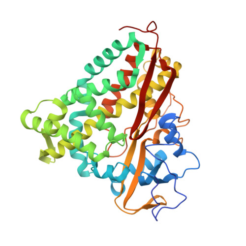
Crystal structure of the carbon monoxide-substrate-cytochrome P-450CAM ternary complex.

Raag, R.Poulos, T.L.

(1989) Biochemistry 28: 7586-7592

  • DOI: https://doi.org/10.1021/bi00445a013
  • Primary Citation Related Structures: 
    3CPP

  • PubMed Abstract: 

    The crystal structure of the ternary complex formed between carbon monoxide (CO), camphor, and ferrous cytochrome P-450CAM has been refined to an R value of 17.9% at 1.9-A resolution. To accommodate the CO molecule, the substrate, camphor, moves about 0.8 A while at the same time remaining in nonbonded contact with CO. The average temperature factor of the camphor atoms is about 50% higher in the CO complex, suggesting that the camphor is more loosely bound in this ternary complex. The Fe-C-O angle is about 166 degrees, and thus, CO appears to be bent from the heme normal, as it is in various CO-globin complexes, due to steric interactions with active site groups. The oxygen atom of the CO molecule is nestled into a groove formed by an unusual helical hydrogen bond in the distal helix between the highly conserved Thr 252 and Gly 248 residues. In the transition from the ferric camphor-bound binary complex to the ferrous CO-camphor-bound ternary complex, the heme iron atom moves into the plane defined by the pyrrole nitrogens by about 0.41 A. Although the axial Cys ligand also moves toward the heme, the S-Fe bond stretches from about 2.20 A in the absence of CO to about 2.41 A once CO has bound.


  • Organizational Affiliation
    • Center for Advanced Research in Biotechnology of the Maryland Biotechnology Institute, University of Maryland, Rockville 20850.

Macromolecule Content 

  • Total Structure Weight: 47.39 kDa 
  • Atom Count: 3,534 
  • Modeled Residue Count: 405 
  • Deposited Residue Count: 414 
  • Unique protein chains: 1

Macromolecules

Find similar proteins by:|  3D Structure
Entity ID: 1
MoleculeChains  Sequence LengthOrganismDetailsImage
CYTOCHROME P450-CAM414Pseudomonas putidaMutation(s): 0 
EC: 1.14.15.1
UniProt
Find proteins for P00183 (Pseudomonas putida)
Explore P00183 
Go to UniProtKB:  P00183
Entity Groups
Sequence Clusters30% Identity50% Identity70% Identity90% Identity95% Identity100% Identity
UniProt GroupP00183
Sequence Annotations
Expand
Reference Sequence

Small Molecules

Ligands 3 Unique
IDChains Name / Formula / InChI Key2D Diagram3D Interactions
HEM

Query on HEM


Download:Ideal Coordinates CCD File
B [auth A]PROTOPORPHYRIN IX CONTAINING FE
C34 H32 Fe N4 O4
KABFMIBPWCXCRK-RGGAHWMASA-L
CAM

Query on CAM


Download:Ideal Coordinates CCD File
D [auth A]CAMPHOR
C10 H16 O
DSSYKIVIOFKYAU-XCBNKYQSSA-N
CMO

Query on CMO


Download:Ideal Coordinates CCD File
C [auth A]CARBON MONOXIDE
C O
UGFAIRIUMAVXCW-UHFFFAOYSA-N

Experimental Data & Validation

Experimental Data

  • Method: X-RAY DIFFRACTION
  • Resolution: 1.90 Å
  • R-Value Observed: 0.190 (Depositor) 
Space Group: P 21 21 21
Unit Cell:
Length ( Å )Angle ( ˚ )
a = 108.67α = 90
b = 103.9β = 90
c = 36.38γ = 90
Software Package:
Software NamePurpose
PROFFTrefinement

Structure Validation

View Full Validation Report


Entry History 

Deposition Data

Revision History  (Full details and data files)

  • Version 1.0: 1990-04-15
    Type: Initial release
  • Version 1.1: 2008-03-25
    Changes: Version format compliance
  • Version 1.2: 2011-07-13
    Changes: Version format compliance
  • Version 1.3: 2017-11-29
    Changes: Derived calculations, Other
  • Version 1.4: 2024-02-21
    Changes: Data collection, Database references, Derived calculations