3COG | pdb_00003cog

Crystal structure of human cystathionase (Cystathionine gamma lyase) in complex with DL-propargylglycine


Experimental Data Snapshot

  • Method: X-RAY DIFFRACTION
  • Resolution: 2.00 Å
  • R-Value Free: 
    0.204 (Depositor), 0.206 (DCC) 
  • R-Value Work: 
    0.158 (Depositor), 0.164 (DCC) 
  • R-Value Observed: 
    0.161 (Depositor) 

Starting Model: experimental
View more details

wwPDB Validation 3D Report Full Report

Validation slider image for 3COG

Ligand Structure Quality Assessment 


This is version 1.2 of the entry. See complete history

Literature

Structural basis for the inhibition mechanism of human cystathionine gamma-lyase, an enzyme responsible for the production of H(2)S.

Sun, Q.Collins, R.Huang, S.Holmberg-Schiavone, L.Anand, G.S.Tan, C.H.van-den-Berg, S.Deng, L.W.Moore, P.K.Karlberg, T.Sivaraman, J.

(2009) J Biological Chem 284: 3076-3085

  • DOI: https://doi.org/10.1074/jbc.M805459200
  • Primary Citation Related Structures: 
    2NMP, 3COG, 3ELP

  • PubMed Abstract: 

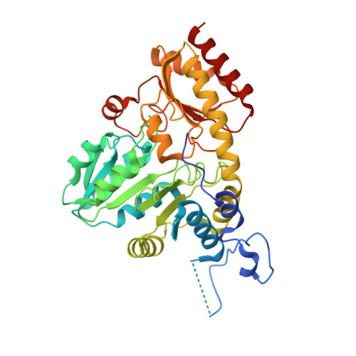
    Impairment of the formation or action of hydrogen sulfide (H(2)S), an endogenous gasotransmitter, is associated with various diseases, such as hypertension, diabetes mellitus, septic and hemorrhagic shock, and pancreatitis. Cystathionine beta-synthase and cystathionine gamma-lyase (CSE) are two pyridoxal-5'-phosphate (PLP)-dependent enzymes largely responsible for the production of H(2)S in mammals. Inhibition of CSE by DL-propargylglycine (PAG) has been shown to alleviate disease symptoms. Here we report crystal structures of human CSE (hCSE), in apo form, and in complex with PLP and PLP.PAG. Structural characterization, combined with biophysical and biochemical studies, provides new insights into the inhibition mechanism of hCSE-mediated production of H(2)S. Transition from the open form of apo-hCSE to the closed PLP-bound form reveals large conformational changes hitherto not reported. In addition, PAG binds hCSE via a unique binding mode, not observed in PAG-enzyme complexes previously. The interaction of PAG-hCSE was not predicted based on existing information from known PAG complexes. The structure of hCSE.PLP.PAG complex highlights the particular importance of Tyr(114) in hCSE and the mechanism of PAG-dependent inhibition of hCSE. These results provide significant insights, which will facilitate the structure-based design of novel inhibitors of hCSE to aid in the development of therapies for diseases involving disorders of sulfur metabolism.


  • Organizational Affiliation
    • Department of Biological Sciences, National University of Singapore, 117543 Singapore.

Macromolecule Content 

  • Total Structure Weight: 179.18 kDa 
  • Atom Count: 13,153 
  • Modeled Residue Count: 1,531 
  • Deposited Residue Count: 1,612 
  • Unique protein chains: 1

Macromolecules

Find similar proteins by:|  3D Structure
Entity ID: 1
MoleculeChains  Sequence LengthOrganismDetailsImage
Cystathionine gamma-lyase
A, B, C, D
403Homo sapiensMutation(s): 0 
Gene Names: CTH
EC: 4.4.1.1 (PDB Primary Data), 4.4.1.2 (UniProt)
UniProt & NIH Common Fund Data Resources
Find proteins for P32929 (Homo sapiens)
Explore P32929 
Go to UniProtKB:  P32929
PHAROS:  P32929
GTEx:  ENSG00000116761 
Entity Groups
Sequence Clusters30% Identity50% Identity70% Identity90% Identity95% Identity100% Identity
UniProt GroupP32929
Sequence Annotations
Expand
Reference Sequence

Small Molecules

Ligands 5 Unique
IDChains Name / Formula / InChI Key2D Diagram3D Interactions
PLP

Query on PLP


Download:Ideal Coordinates CCD File
F [auth A],
K [auth C],
O [auth D]
PYRIDOXAL-5'-PHOSPHATE
C8 H10 N O6 P
NGVDGCNFYWLIFO-UHFFFAOYSA-N
2AG

Query on 2AG


Download:Ideal Coordinates CCD File
G [auth A],
L [auth C],
P [auth D]
(2S)-2-aminopent-4-enoic acid
C5 H9 N O2
WNNNWFKQCKFSDK-BYPYZUCNSA-N
PEG

Query on PEG


Download:Ideal Coordinates CCD File
I [auth A]DI(HYDROXYETHYL)ETHER
C4 H10 O3
MTHSVFCYNBDYFN-UHFFFAOYSA-N
GOL

Query on GOL


Download:Ideal Coordinates CCD File
H [auth A],
M [auth C],
Q [auth D],
R [auth D],
S [auth D]
GLYCEROL
C3 H8 O3
PEDCQBHIVMGVHV-UHFFFAOYSA-N
NO3

Query on NO3


Download:Ideal Coordinates CCD File
E [auth A],
J [auth C],
N [auth D]
NITRATE ION
N O3
NHNBFGGVMKEFGY-UHFFFAOYSA-N

Experimental Data & Validation

Experimental Data

  • Method: X-RAY DIFFRACTION
  • Resolution: 2.00 Å
  • R-Value Free:  0.204 (Depositor), 0.206 (DCC) 
  • R-Value Work:  0.158 (Depositor), 0.164 (DCC) 
  • R-Value Observed: 0.161 (Depositor) 
Space Group: P 21 21 21
Unit Cell:
Length ( Å )Angle ( ˚ )
a = 105.35α = 90
b = 107.22β = 90
c = 153.31γ = 90
Software Package:
Software NamePurpose
REFMACrefinement
MxCuBEdata collection
XDSdata reduction
XSCALEdata scaling
MOLREPphasing

Structure Validation

View Full Validation Report


Ligand Structure Quality Assessment 


Entry History 

Revision History  (Full details and data files)

  • Version 1.0: 2008-05-27
    Type: Initial release
  • Version 1.1: 2011-07-13
    Changes: Non-polymer description, Version format compliance
  • Version 1.2: 2023-08-30
    Changes: Data collection, Database references, Derived calculations, Refinement description