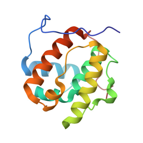
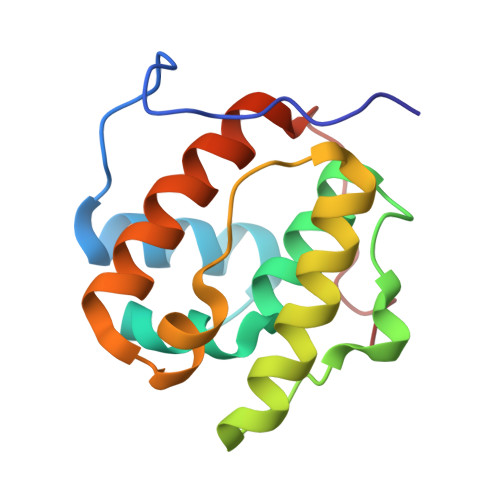
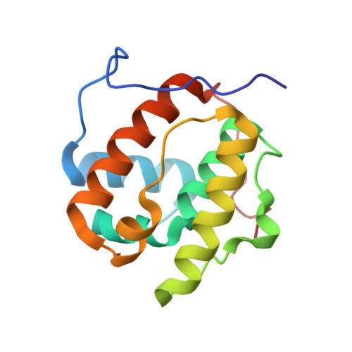
3CO1

Crystal structure of microtubule binding domain of human EB3


Experimental Data Snapshot

  • Method: X-RAY DIFFRACTION
  • Resolution: 1.40 Å
  • R-Value Free: 0.234 
  • R-Value Work: 0.200 
  • R-Value Observed: 0.202 

Starting Model: experimental
View more details

wwPDB Validation   3D Report Full Report


This is version 1.2 of the entry. See complete history


Literature

Mammalian end binding proteins control persistent microtubule growth.

Komarova, Y.De Groot, C.O.Grigoriev, I.Gouveia, S.M.Munteanu, E.L.Schober, J.M.Honnappa, S.Buey, R.M.Hoogenraad, C.C.Dogterom, M.Borisy, G.G.Steinmetz, M.O.Akhmanova, A.

(2009) J Cell Biol 

  • DOI: https://doi.org/10.1083/jcb.200807179
  • Primary Citation of Related Structures:  
    3CO1

  • PubMed Abstract: 

    End binding proteins (EBs) are highly conserved core components of microtubule plus-end tracking protein networks. Here we investigated the roles of the three mammalian EBs in controlling microtubule dynamics and analyzed the domains involved. Protein depletion and rescue experiments showed that EB1 and EB3, but not EB2, promote persistent microtubule growth by suppressing catastrophes. Furthermore, we demonstrated in vitro and in cells that the EB plus-end tracking behavior depends on the calponin homology domain but does not require dimer formation. In contrast, dimerization is necessary for the EB anti-catastrophe activity in cells; this explains why the EB1 dimerization domain, which disrupts native EB dimers, exhibits a dominant-negative effect. When microtubule dynamics is reconstituted with purified tubulin, EBs promote rather than inhibit catastrophes, suggesting that in cells EBs prevent catastrophes by counteracting other microtubule regulators. This probably occurs through their action on microtubule ends, because catastrophe suppression does not require the EB domains needed for binding to known EB partners.


  • Organizational Affiliation

    Department of Cell and Molecular Biology, Northwestern University Medical School, Chicago, IL 60611, USA.


Macromolecules
Find similar proteins by:  (by identity cutoff)  |  3D Structure
Entity ID: 1
MoleculeChains Sequence LengthOrganismDetailsImage
Microtubule-associated protein RP/EB family member 3132Homo sapiensMutation(s): 0 
UniProt & NIH Common Fund Data Resources
Find proteins for Q9UPY8 (Homo sapiens)
Explore Q9UPY8 
Go to UniProtKB:  Q9UPY8
PHAROS:  Q9UPY8
GTEx:  ENSG00000084764 
Entity Groups  
Sequence Clusters30% Identity50% Identity70% Identity90% Identity95% Identity100% Identity
UniProt GroupQ9UPY8
Sequence Annotations
Expand
  • Reference Sequence
Experimental Data & Validation

Experimental Data

  • Method: X-RAY DIFFRACTION
  • Resolution: 1.40 Å
  • R-Value Free: 0.234 
  • R-Value Work: 0.200 
  • R-Value Observed: 0.202 
  • Space Group: P 21 21 2
Unit Cell:
Length ( Å )Angle ( ˚ )
a = 47.188α = 90
b = 85.365β = 90
c = 32.161γ = 90
Software Package:
Software NamePurpose
REFMACrefinement
XDSdata scaling
XDSdata reduction
XSCALEdata scaling
PHASERphasing

Structure Validation

View Full Validation Report


Entry History 

Deposition Data

Revision History  (Full details and data files)

  • Version 1.0: 2009-03-17
    Type: Initial release
  • Version 1.1: 2011-07-13
    Changes: Advisory, Version format compliance
  • Version 1.2: 2023-11-01
    Changes: Data collection, Database references, Refinement description