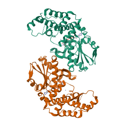
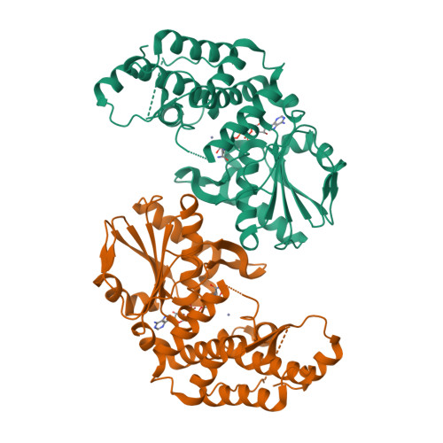
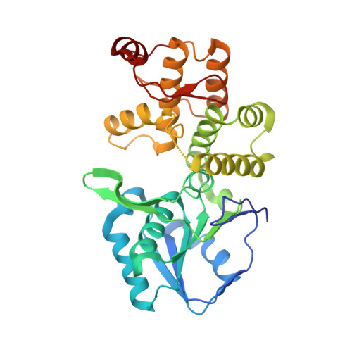
3CLH

Crystal structure of 3-dehydroquinate synthase (DHQS)from Helicobacter pylori


Experimental Data Snapshot

  • Method: X-RAY DIFFRACTION
  • Resolution: 2.40 Å
  • R-Value Free: 0.258 
  • R-Value Work: 0.207 

Starting Model: experimental
View more details

wwPDB Validation   3D Report Full Report


This is version 1.2 of the entry. See complete history


Literature

Structure-based inhibitor discovery of Helicobacter pylori dehydroquinate synthase.

Liu, J.S.Cheng, W.C.Wang, H.J.Chen, Y.C.Wang, W.C.

(2008) Biochem Biophys Res Commun 373: 1-7

  • DOI: https://doi.org/10.1016/j.bbrc.2008.05.070
  • Primary Citation of Related Structures:  
    3CLH

  • PubMed Abstract: 

    Dehydroquinate synthase (DHQS) is a nicotinamide adenine dinucleotide (NAD)-dependent enzyme that converts 3-deoxy-D-arabino-heptulosonate 7-phosphate (DAHP) into 3-dehydroquinate (DHQ). Since it catalyzes the second key step in the shikimate pathway, which is crucial for the aromatic amino acid metabolism in bacteria, fungi, and plants, but not in mammals, DHQS is a potential target for new antimicrobial agents, anti-parasitic agents and herbicides. The crystal structure of Helicobacter pylori DHQS (HpDHQS) complexed with NAD has been determined at 2.4-A resolution and was found to possess an N-terminal Rossmann-fold domain and a C-terminal alpha-helical domain. Structural comparison reveals that the binary complex adopts an open-state conformation and shares conserved residues in the binding pocket. Virtual docking of compounds into the active site of the HpDHQS structure using the GOLD docking program led to the identification of several inhibitors. The most active compound had an IC(50) value of 61 microM, which may serve as a lead for potent inhibitors.


  • Organizational Affiliation

    Institute of Molecular and Cellular Biology and Department of Life Sciences, National Tsing Hua University, 101, Section 2, Kuang-Fu Road, Hsinchu 300, Taiwan.


Macromolecules
Find similar proteins by:  (by identity cutoff)  |  3D Structure
Entity ID: 1
MoleculeChains Sequence LengthOrganismDetailsImage
3-dehydroquinate synthase
A, B
343Helicobacter pyloriMutation(s): 0 
Gene Names: aroB
EC: 4.2.3.4
UniProt
Find proteins for P56081 (Helicobacter pylori (strain ATCC 700392 / 26695))
Explore P56081 
Go to UniProtKB:  P56081
Entity Groups  
Sequence Clusters30% Identity50% Identity70% Identity90% Identity95% Identity100% Identity
UniProt GroupP56081
Sequence Annotations
Expand
  • Reference Sequence
Experimental Data & Validation

Experimental Data

  • Method: X-RAY DIFFRACTION
  • Resolution: 2.40 Å
  • R-Value Free: 0.258 
  • R-Value Work: 0.207 
  • Space Group: H 3
Unit Cell:
Length ( Å )Angle ( ˚ )
a = 158.294α = 90
b = 158.294β = 90
c = 97.387γ = 120
Software Package:
Software NamePurpose
CrystalCleardata collection
AMoREphasing
REFMACrefinement
DENZOdata reduction
SCALEPACKdata scaling

Structure Validation

View Full Validation Report


Entry History 

Deposition Data

Revision History  (Full details and data files)

  • Version 1.0: 2009-03-24
    Type: Initial release
  • Version 1.1: 2011-07-13
    Changes: Version format compliance
  • Version 1.2: 2023-11-01
    Changes: Data collection, Database references, Derived calculations, Refinement description