Refined crystal structure of type III chloramphenicol acetyltransferase at 1.75 A resolution.
Leslie, A.G.(1990) J Mol Biol 213: 167-186
- PubMed: 2187098 Search on PubMed
- DOI: https://doi.org/10.1016/S0022-2836(05)80129-9
- Primary Citation Related Structures:
3CLA - PubMed Abstract:
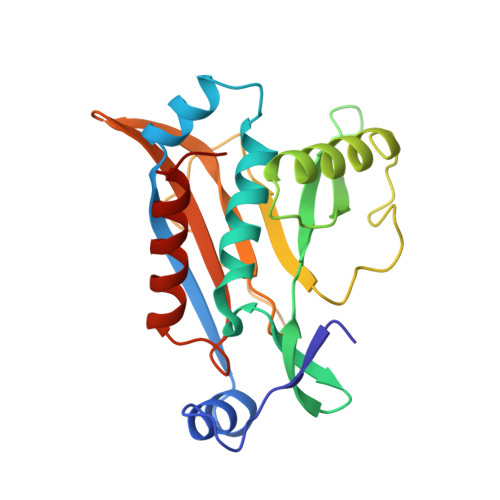
High level bacterial resistance to chloramphenicol is generally due to O-acetylation of the antibiotic in a reaction catalysed by chloramphenicol acetyltransferase (CAT, EC 2.3.1.28) in which acetyl-coenzyme A is the acyl donor. The crystal structure of the type III enzyme from Escherichia coli with chloramphenicol bound has been determined and refined at 1.75 A resolution, using a restrained parameter reciprocal space least squares procedure. The refined model, which includes chloramphenicol, 204 solvent molecules and two cobalt ions has a crystallographic R-factor of 18.3% for 27,300 reflections between 6 and 1.75 A resolution. The root-mean-square deviation in bond lengths from ideal values is 0.02 A. The cobalt ions play a crucial role in stabilizing the packing of the molecule in the crystal lattice. CAT is a trimer of identical subunits (monomer Mr 25,000) and the trimeric structure is stabilized by a number of hydrogen bonds, some of which result in the extension of a beta-sheet across the subunit interface. Chloramphenicol binds in a deep pocket located at the boundary between adjacent subunits of the trimer, such that the majority of residues forming the binding pocket belong to one subunit while the catalytically essential histidine belongs to the adjacent subunit. His195 is appropriately positioned to act as a general base catalyst in the reaction, and the required tautomeric stabilization is provided by an unusual interaction with a main-chain carbonyl oxygen.
- Blackett Laboratory, Imperial College, London, U.K.
Organizational Affiliation: