3CL9 | pdb_00003cl9

Structure of bifunctional TcDHFR-TS in complex with MTX


Experimental Data Snapshot

  • Method: X-RAY DIFFRACTION
  • Resolution: 3.30 Å
  • R-Value Free: 
    0.249 (Depositor), 0.241 (DCC) 
  • R-Value Work: 
    0.206 (Depositor), 0.199 (DCC) 

Starting Model: experimental
View more details

wwPDB Validation 3D Report Full Report

Validation slider image for 3CL9

Ligand Structure Quality Assessment 


This is version 1.2 of the entry. See complete history

Literature

Structure-based approach to pharmacophore identification, in silico screening, and three-dimensional quantitative structure-activity relationship studies for inhibitors of Trypanosoma cruzi dihydrofolate reductase function.

Schormann, N.Senkovich, O.Walker, K.Wright, D.L.Anderson, A.C.Rosowsky, A.Ananthan, S.Shinkre, B.Velu, S.Chattopadhyay, D.

(2008) Proteins 73: 889-901

  • DOI: https://doi.org/10.1002/prot.22115
  • Primary Citation Related Structures: 
    2H2Q, 3CL9, 3CLB

  • PubMed Abstract: 

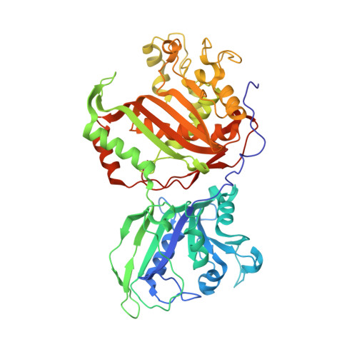
    We have employed a structure-based three-dimensional quantitative structure-activity relationship (3D-QSAR) approach to predict the biochemical activity for inhibitors of T. cruzi dihydrofolate reductase-thymidylate synthase (DHFR-TS). Crystal structures of complexes of the enzyme with eight different inhibitors of the DHFR activity together with the structure in the substrate-free state (DHFR domain) were used to validate and refine docking poses of ligands that constitute likely active conformations. Structural information from these complexes formed the basis for the structure-based alignment used as input for the QSAR study. Contrary to indirect ligand-based approaches the strategy described here employs a direct receptor-based approach. The goal is to generate a library of selective lead inhibitors for further development as antiparasitic agents. 3D-QSAR models were obtained for T. cruzi DHFR-TS (30 inhibitors in learning set) and human DHFR (36 inhibitors in learning set) that show a very good agreement between experimental and predicted enzyme inhibition data. For crossvalidation of the QSAR model(s), we have used the 10% leave-one-out method. The derived 3D-QSAR models were tested against a few selected compounds (a small test set of six inhibitors for each enzyme) with known activity, which were not part of the learning set, and the quality of prediction of the initial 3D-QSAR models demonstrated that such studies are feasible. Further refinement of the models through integration of additional activity data and optimization of reliable docking poses is expected to lead to an improved predictive ability.


  • Organizational Affiliation
    • Department of Pharmaceutical Sciences, University of Alabama at Birmingham, Birmingham, Alabama 35294, USA.

Macromolecule Content 

  • Total Structure Weight: 60.83 kDa 
  • Atom Count: 4,157 
  • Modeled Residue Count: 505 
  • Deposited Residue Count: 521 
  • Unique protein chains: 1

Macromolecules

Find similar proteins by:|  3D Structure
Entity ID: 1
MoleculeChains  Sequence LengthOrganismDetailsImage
Bifunctional dihydrofolate reductase-thymidylate synthase (DHFR-TS)521Trypanosoma cruziMutation(s): 0 
EC: 1.5.1.3 (PDB Primary Data), 2.1.1.45 (PDB Primary Data)
UniProt
Find proteins for Q27793 (Trypanosoma cruzi)
Explore Q27793 
Go to UniProtKB:  Q27793
Entity Groups
Sequence Clusters30% Identity50% Identity70% Identity90% Identity95% Identity100% Identity
UniProt GroupQ27793
Sequence Annotations
Expand
Reference Sequence

Small Molecules

Ligands 6 Unique
IDChains Name / Formula / InChI Key2D Diagram3D Interactions
NAP

Query on NAP


Download:Ideal Coordinates CCD File
G [auth A]NADP NICOTINAMIDE-ADENINE-DINUCLEOTIDE PHOSPHATE
C21 H28 N7 O17 P3
XJLXINKUBYWONI-NNYOXOHSSA-N
MTX

Query on MTX


Download:Ideal Coordinates CCD File
H [auth A]METHOTREXATE
C20 H22 N8 O5
FBOZXECLQNJBKD-ZDUSSCGKSA-N
UMP

Query on UMP


Download:Ideal Coordinates CCD File
I [auth A]2'-DEOXYURIDINE 5'-MONOPHOSPHATE
C9 H13 N2 O8 P
JSRLJPSBLDHEIO-SHYZEUOFSA-N
SO4

Query on SO4


Download:Ideal Coordinates CCD File
B [auth A],
C [auth A]
SULFATE ION
O4 S
QAOWNCQODCNURD-UHFFFAOYSA-L
EDO

Query on EDO


Download:Ideal Coordinates CCD File
J [auth A],
K [auth A]
1,2-ETHANEDIOL
C2 H6 O2
LYCAIKOWRPUZTN-UHFFFAOYSA-N
CL

Query on CL


Download:Ideal Coordinates CCD File
D [auth A],
E [auth A],
F [auth A]
CHLORIDE ION
Cl
VEXZGXHMUGYJMC-UHFFFAOYSA-M

Experimental Data & Validation

Experimental Data

  • Method: X-RAY DIFFRACTION
  • Resolution: 3.30 Å
  • R-Value Free:  0.249 (Depositor), 0.241 (DCC) 
  • R-Value Work:  0.206 (Depositor), 0.199 (DCC) 
Space Group: I 41 2 2
Unit Cell:
Length ( Å )Angle ( ˚ )
a = 121.05α = 90
b = 121.05β = 90
c = 260.76γ = 90
Software Package:
Software NamePurpose
REFMACrefinement
HKL-2000data collection
DENZOdata reduction
SCALEPACKdata scaling
PHASERphasing

Structure Validation

View Full Validation Report


Ligand Structure Quality Assessment 


Entry History 

Deposition Data

Revision History  (Full details and data files)

  • Version 1.0: 2009-01-06
    Type: Initial release
  • Version 1.1: 2011-07-13
    Changes: Advisory, Version format compliance
  • Version 1.2: 2023-08-30
    Changes: Data collection, Database references, Derived calculations, Refinement description