3CDY | pdb_00003cdy

AL-09 H87Y, immunoglobulin light chain variable domain


Experimental Data Snapshot

  • Method: X-RAY DIFFRACTION
  • Resolution: 2.43 Å
  • R-Value Free: 
    0.263 (Depositor), 0.263 (DCC) 
  • R-Value Work: 
    0.189 (Depositor), 0.196 (DCC) 
  • R-Value Observed: 
    0.193 (Depositor) 

Starting Model: experimental
View more details

wwPDB Validation 3D Report Full Report

Validation slider image for 3CDY

This is version 1.3 of the entry. See complete history

Literature

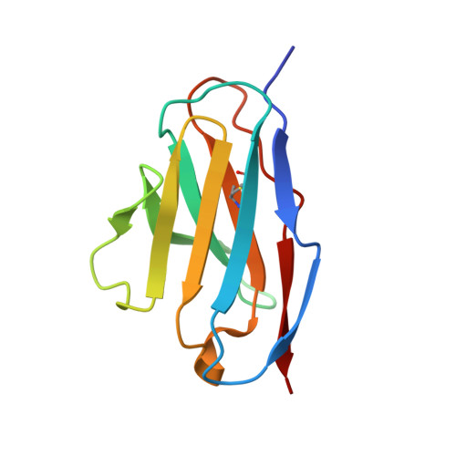
Structural insights into the role of mutations in amyloidogenesis.

Baden, E.M.Randles, E.G.Aboagye, A.K.Thompson, J.R.Ramirez-Alvarado, M.

(2008) J Biological Chem 283: 30950-30956

  • DOI: https://doi.org/10.1074/jbc.M804822200
  • Primary Citation Related Structures: 
    3CDC, 3CDF, 3CDY

  • PubMed Abstract: 

    Mechanisms of amyloidogenesis are not well understood, including potential structural contributions of mutations in the process. Our previous research indicated that the dimer interface of amyloidogenic immunoglobulin light chain protein AL-09 is twisted 90 degrees relative to the protein from its germline sequence, kappaI O18/O8. Here we report a systematic restoration of AL-09 to its germline sequence by mutating the non-conservative somatic mutations located in the light chain dimer interface. Among these mutants, we find a correlation between increased thermodynamic stability and an increase in the lag time for fibril formation. The restorative mutant AL-09 H87Y completes the trifecta and restores the dimer interface observed in kappaI O18/O8, emphasizing the potential importance of the structural integrity of these proteins to protect against amyloidogenicity. We also find that adding amyloidogenic mutations into the germline protein illustrates mutational cooperativity in promoting amyloidogenesis.


  • Organizational Affiliation
    • Department of Biochemistry, College of Medicine, Mayo Clinic, Rochester, Minnesota 55905, USA.

Macromolecule Content 

  • Total Structure Weight: 23.99 kDa 
  • Atom Count: 1,785 
  • Modeled Residue Count: 216 
  • Deposited Residue Count: 218 
  • Unique protein chains: 1

Macromolecules

Find similar proteins by:|  3D Structure
Entity ID: 1
MoleculeChains  Sequence LengthOrganismDetailsImage
IMMUNOGLOBULIN LIGHT CHAIN
A, B
109Homo sapiensMutation(s): 0 
Gene Names: kI O18/O8 germline
UniProt & NIH Common Fund Data Resources
Find proteins for P01594 (Homo sapiens)
Explore P01594 
Go to UniProtKB:  P01594
GTEx:  ENSG00000242076 
Entity Groups
Sequence Clusters30% Identity50% Identity70% Identity90% Identity95% Identity100% Identity
UniProt GroupP01594
Sequence Annotations
Expand
Reference Sequence

Experimental Data & Validation

Experimental Data

  • Method: X-RAY DIFFRACTION
  • Resolution: 2.43 Å
  • R-Value Free:  0.263 (Depositor), 0.263 (DCC) 
  • R-Value Work:  0.189 (Depositor), 0.196 (DCC) 
  • R-Value Observed: 0.193 (Depositor) 
Space Group: P 61
Unit Cell:
Length ( Å )Angle ( ˚ )
a = 73.971α = 90
b = 73.971β = 90
c = 95.07γ = 120
Software Package:
Software NamePurpose
REFMACrefinement
HKL-2000data collection
HKL-2000data reduction
SCALEPACKdata scaling
PHASERphasing

Structure Validation

View Full Validation Report


Entry History 

Deposition Data

Revision History  (Full details and data files)

  • Version 1.0: 2008-09-02
    Type: Initial release
  • Version 1.1: 2011-07-13
    Changes: Advisory, Version format compliance
  • Version 1.2: 2023-08-30
    Changes: Data collection, Database references, Refinement description
  • Version 1.3: 2024-11-06
    Changes: Structure summary