3CAA | pdb_00003caa

CLEAVED ANTICHYMOTRYPSIN A347R


Experimental Data Snapshot

  • Method: X-RAY DIFFRACTION
  • Resolution: 2.40 Å
  • R-Value Free: 
    0.280 (Depositor) 
  • R-Value Work: 
    0.190 (Depositor) 
  • R-Value Observed: 
    0.190 (Depositor) 

Starting Model: experimental
View more details

wwPDB Validation 3D Report Full Report

Validation slider image for 3CAA

This is version 1.5 of the entry. See complete history

Literature

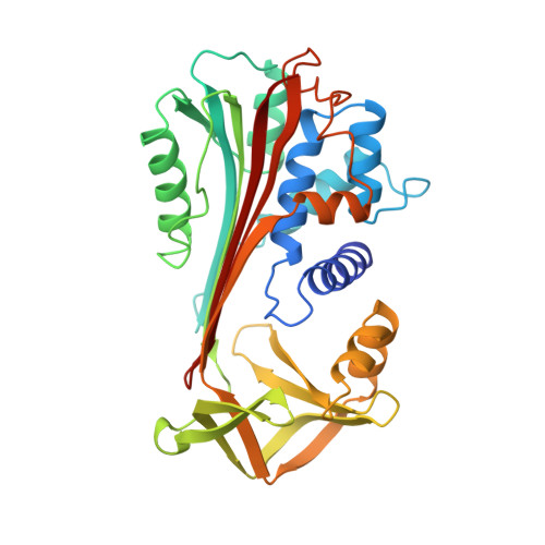
Engineering an anion-binding cavity in antichymotrypsin modulates the "spring-loaded" serpin-protease interaction.

Lukacs, C.M.Rubin, H.Christianson, D.W.

(1998) Biochemistry 37: 3297-3304

  • DOI: https://doi.org/10.1021/bi972359e
  • Primary Citation Related Structures: 
    1AS4, 3CAA, 4CAA

  • PubMed Abstract: 

    Expressed in a kinetically trapped folding state, a serpin couples the thermodynamic driving force of a massive beta-sheet rearrangement to the inhibition of a target protease. Hence, the serpin-protease interaction is the premier example of a "spring-loaded" protein-protein interaction. Amino acid substitutions in the hinge region of a serpin reactive loop can weaken the molecular spring, which converts the serpin from an inhibitor into a substrate. To probe the molecular basis of this conversion, we report the crystal structure of A349R antichymotrypsin in the reactive loop cleaved state at 2.1 A resolution. This amino acid substitution does not block the beta-sheet rearrangement despite the burial of R349 in the hydrophobic core of the cleaved serpin along with a salt-linked acetate ion. The inhibitory activity of this serpin variant is not obliterated; remarkably, its inhibitory properties are anion-dependent due to the creation of an anion-binding cavity in the cleaved serpin.


  • Organizational Affiliation
    • Department of Chemistry, University of Pennsylvania, Philadelphia 19104, USA.

Macromolecule Content 

  • Total Structure Weight: 42.97 kDa 
  • Atom Count: 2,978 
  • Modeled Residue Count: 366 
  • Deposited Residue Count: 378 
  • Unique protein chains: 2

Macromolecules

Find similar proteins by:|  3D Structure
Entity ID: 1
MoleculeChains  Sequence LengthOrganismDetailsImage
ANTICHYMOTRYPSIN341Homo sapiensMutation(s): 1 
Gene Names: ACT
UniProt & NIH Common Fund Data Resources
Find proteins for P01011 (Homo sapiens)
Explore P01011 
Go to UniProtKB:  P01011
PHAROS:  P01011
GTEx:  ENSG00000196136 
Entity Groups
Sequence Clusters30% Identity50% Identity70% Identity90% Identity95% Identity100% Identity
UniProt GroupP01011
Sequence Annotations
Expand
Reference Sequence
Find similar proteins by:|  3D Structure
Entity ID: 2
MoleculeChains  Sequence LengthOrganismDetailsImage
ANTICHYMOTRYPSIN37Homo sapiensMutation(s): 0 
Gene Names: ACT
UniProt & NIH Common Fund Data Resources
Find proteins for P01011 (Homo sapiens)
Explore P01011 
Go to UniProtKB:  P01011
PHAROS:  P01011
GTEx:  ENSG00000196136 
Entity Groups
Sequence Clusters30% Identity50% Identity70% Identity90% Identity95% Identity100% Identity
UniProt GroupP01011
Sequence Annotations
Expand
Reference Sequence

Experimental Data & Validation

Experimental Data

  • Method: X-RAY DIFFRACTION
  • Resolution: 2.40 Å
  • R-Value Free:  0.280 (Depositor) 
  • R-Value Work:  0.190 (Depositor) 
  • R-Value Observed: 0.190 (Depositor) 
Space Group: P 21 21 21
Unit Cell:
Length ( Å )Angle ( ˚ )
a = 74.12α = 90
b = 77.73β = 90
c = 79.49γ = 90
Software Package:
Software NamePurpose
X-PLORmodel building
X-PLORrefinement
DENZOdata reduction
SCALEPACKdata scaling
X-PLORphasing

Structure Validation

View Full Validation Report


Entry History 

Deposition Data

Revision History  (Full details and data files)

  • Version 1.0: 1998-02-25
    Type: Initial release
  • Version 1.1: 2008-03-04
    Changes: Version format compliance
  • Version 1.2: 2011-07-13
    Changes: Version format compliance
  • Version 1.3: 2021-11-03
    Changes: Database references, Other
  • Version 1.4: 2023-08-09
    Changes: Refinement description
  • Version 1.5: 2024-05-22
    Changes: Data collection