3CA9 | pdb_00003ca9

Evolution of chlorella virus dUTPase


Experimental Data Snapshot

  • Method: X-RAY DIFFRACTION
  • Resolution: 3.00 Å
  • R-Value Free: 
    0.307 (Depositor), 0.219 (DCC) 
  • R-Value Work: 
    0.232 (Depositor), 0.224 (DCC) 
  • R-Value Observed: 
    0.232 (Depositor) 

wwPDB Validation 3D Report Full Report

Validation slider image for 3CA9

Ligand Structure Quality Assessment 


This is version 1.2 of the entry. See complete history

Literature

Crystallization and crystal-packing studies of Chlorella virus deoxyuridine triphosphatase.

Homma, K.Moriyama, H.

(2009) Acta Crystallogr Sect F Struct Biol Cryst Commun 65: 1030-1034

  • DOI: https://doi.org/10.1107/S1744309109034459
  • Primary Citation Related Structures: 
    3C2T, 3C3I, 3CA9

  • PubMed Abstract: 

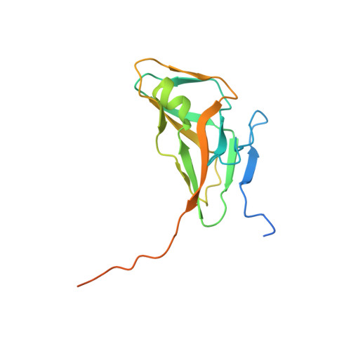
    The 141-amino-acid deoxyuridine triphosphatase (dUTPase) from the algal Chlorella virus IL-3A and its Glu81Ser/Thr84Arg-mutant derivative Mu-22 were crystallized using the hanging-drop vapor-diffusion method at 298 K with polyethylene glycol as the precipitant. An apo IL-3A dUTPase with an amino-terminal T7 epitope tag and a carboxy-terminal histidine tag yielded cubic P2(1)3 crystals with unit-cell parameter a = 106.65 A. In the presence of dUDP, the enzyme produced thin stacked orthorhombic P222 crystals with unit-cell parameters a = 81.0, b = 96.2, c = 132.8 A. T7-histidine-tagged Mu-22 dUTPase formed thin stacked rectangular crystals. Amino-terminal histidine-tagged dUTPases did not crystallize but formed aggregates. Glycyl-seryl-tagged dUTPases yielded cubic P2(1)3 IL-3A crystals with unit-cell parameter a = 105.68 A and hexagonal P6(3) Mu-22 crystals with unit-cell parameters a = 132.07, c = 53.45 A, gamma = 120 degrees . Owing to the Thr84Arg mutation, Mu-22 dUTPase had different monomer-to-monomer interactions to those of IL-3A dUTPase.


  • Organizational Affiliation
    • Department of Chemistry, University of Nebraska-Lincoln, NE 68583-0304, USA.

Macromolecule Content 

  • Total Structure Weight: 35.9 kDa 
  • Atom Count: 1,976 
  • Modeled Residue Count: 254 
  • Deposited Residue Count: 330 
  • Unique protein chains: 1

Macromolecules

Find similar proteins by:|  3D Structure
Entity ID: 1
MoleculeChains  Sequence LengthOrganismDetailsImage
Deoxyuridine triphosphatase
A, B
165Paramecium bursaria Chlorella virus IL3AMutation(s): 0 
EC: 3.6.1.23
UniProt
Find proteins for Q5I3E5 (Paramecium bursaria Chlorella virus IL3A)
Explore Q5I3E5 
Go to UniProtKB:  Q5I3E5
Entity Groups
Sequence Clusters30% Identity50% Identity70% Identity90% Identity95% Identity100% Identity
UniProt GroupQ5I3E5
Sequence Annotations
Expand
Reference Sequence

Experimental Data & Validation

Experimental Data

  • Method: X-RAY DIFFRACTION
  • Resolution: 3.00 Å
  • R-Value Free:  0.307 (Depositor), 0.219 (DCC) 
  • R-Value Work:  0.232 (Depositor), 0.224 (DCC) 
  • R-Value Observed: 0.232 (Depositor) 
Space Group: P 21 3
Unit Cell:
Length ( Å )Angle ( ˚ )
a = 105.647α = 90
b = 105.647β = 90
c = 105.647γ = 90
Software Package:
Software NamePurpose
CNSrefinement
CrystalCleardata collection
HKL-2000data reduction
HKL-2000data scaling
CNSphasing

Structure Validation

View Full Validation Report


Ligand Structure Quality Assessment 


Entry History 

Deposition Data

Revision History  (Full details and data files)

  • Version 1.0: 2009-03-03
    Type: Initial release
  • Version 1.1: 2011-07-13
    Changes: Version format compliance
  • Version 1.2: 2024-02-21
    Changes: Data collection, Database references, Derived calculations