3C99 | pdb_00003c99

Structural Basis of Histone H4 Recognition by p55


Experimental Data Snapshot

  • Method: X-RAY DIFFRACTION
  • Resolution: 2.90 Å
  • R-Value Free: 
    0.256 (Depositor), 0.257 (DCC) 
  • R-Value Work: 
    0.219 (Depositor), 0.219 (DCC) 

Starting Model: experimental
View more details

wwPDB Validation 3D Report Full Report

Validation slider image for 3C99

This is version 1.3 of the entry. See complete history

Literature

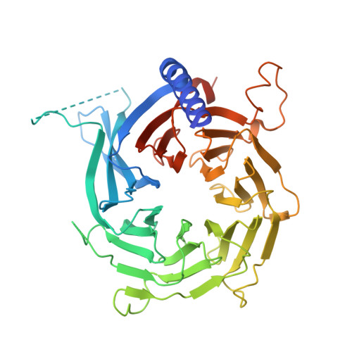
Structural basis of histone H4 recognition by p55.

Song, J.J.Garlick, J.D.Kingston, R.E.

(2008) Genes Dev 22: 1313-1318

  • DOI: https://doi.org/10.1101/gad.1653308
  • Primary Citation Related Structures: 
    3C99, 3C9C

  • PubMed Abstract: 

    p55 is a common component of many chromatin-modifying complexes and has been shown to bind to histones. Here, we present a crystal structure of Drosophila p55 bound to a histone H4 peptide. p55, a predicted WD40 repeat protein, recognizes the first helix of histone H4 via a binding pocket located on the side of a beta-propeller structure. The pocket cannot accommodate the histone fold of H4, which must be altered to allow p55 binding. Reconstitution experiments show that the binding pocket is important to the function of p55-containing complexes. These data demonstrate that WD40 repeat proteins use various surfaces to direct the modification of histones.


  • Organizational Affiliation
    • Department of Molecular Biology, Massachusetts General Hospital, Boston, Massachusetts 02114, USA.

Macromolecule Content 

  • Total Structure Weight: 49.83 kDa 
  • Atom Count: 3,063 
  • Modeled Residue Count: 380 
  • Deposited Residue Count: 432 
  • Unique protein chains: 1

Macromolecules

Find similar proteins by:|  3D Structure
Entity ID: 1
MoleculeChains  Sequence LengthOrganismDetailsImage
Chromatin assembly factor 1 p55 subunit432Drosophila melanogasterMutation(s): 0 
Gene Names: Caf1
UniProt
Find proteins for Q24572 (Drosophila melanogaster)
Explore Q24572 
Go to UniProtKB:  Q24572
Entity Groups
Sequence Clusters30% Identity50% Identity70% Identity90% Identity95% Identity100% Identity
UniProt GroupQ24572
Sequence Annotations
Expand
Reference Sequence

Small Molecules

Ligands 1 Unique
IDChains Name / Formula / InChI Key2D Diagram3D Interactions
CD

Query on CD


Download:Ideal Coordinates CCD File
B [auth A]
C [auth A]
D [auth A]
E [auth A]
F [auth A]
B [auth A],
C [auth A],
D [auth A],
E [auth A],
F [auth A],
G [auth A],
H [auth A],
I [auth A],
J [auth A]
CADMIUM ION
Cd
WLZRMCYVCSSEQC-UHFFFAOYSA-N

Experimental Data & Validation

Experimental Data

  • Method: X-RAY DIFFRACTION
  • Resolution: 2.90 Å
  • R-Value Free:  0.256 (Depositor), 0.257 (DCC) 
  • R-Value Work:  0.219 (Depositor), 0.219 (DCC) 
Space Group: P 31 2 1
Unit Cell:
Length ( Å )Angle ( ˚ )
a = 151.121α = 90
b = 151.121β = 90
c = 98.013γ = 120
Software Package:
Software NamePurpose
HKL-2000data collection
PHASESphasing
CNSrefinement
HKL-2000data reduction
HKL-2000data scaling

Structure Validation

View Full Validation Report


Entry History 

Deposition Data

Revision History  (Full details and data files)

  • Version 1.0: 2008-05-13
    Type: Initial release
  • Version 1.1: 2011-07-13
    Changes: Version format compliance
  • Version 1.2: 2024-02-21
    Changes: Data collection, Database references, Derived calculations
  • Version 1.3: 2024-04-03
    Changes: Refinement description