3C0O

Crystal structure of the proaerolysin mutant Y221G complexed with mannose-6-phosphate


Experimental Data Snapshot

  • Method: X-RAY DIFFRACTION
  • Resolution: 2.50 Å
  • R-Value Free: 0.274 
  • R-Value Work: 0.203 
  • R-Value Observed: 0.207 

Starting Model: experimental
View more details

wwPDB Validation   3D Report Full Report


Ligand Structure Quality Assessment 


This is version 1.5 of the entry. See complete history


Literature

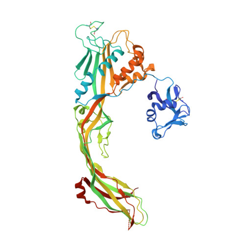
Molecular assembly of the aerolysin pore reveals a swirling membrane-insertion mechanism.

Degiacomi, M.T.Iacovache, I.Pernot, L.Chami, M.Kudryashev, M.Stahlberg, H.van der Goot, F.G.Dal Peraro, M.

(2013) Nat Chem Biol 9: 623-629

  • DOI: https://doi.org/10.1038/nchembio.1312
  • Primary Citation of Related Structures:  
    3C0M, 3C0N, 3C0O

  • PubMed Abstract: 

    Aerolysin is the founding member of a superfamily of β-pore-forming toxins whose pore structure is unknown. We have combined X-ray crystallography, cryo-EM, molecular dynamics and computational modeling to determine the structures of aerolysin mutants in their monomeric and heptameric forms, trapped at various stages of the pore formation process. A dynamic modeling approach based on swarm intelligence was applied, whereby the intrinsic flexibility of aerolysin extracted from new X-ray structures was used to fully exploit the cryo-EM spatial restraints. Using this integrated strategy, we obtained a radically new arrangement of the prepore conformation and a near-atomistic structure of the aerolysin pore, which is fully consistent with all of the biochemical data available so far. Upon transition from the prepore to pore, the aerolysin heptamer shows a unique concerted swirling movement, accompanied by a vertical collapse of the complex, ultimately leading to the insertion of a transmembrane β-barrel.


  • Organizational Affiliation

    1] Institute of Bioengineering, School of Life Sciences, Ecole Polytechnique Fédérale de Lausanne, Lausanne, Switzerland. [2] [3].


Macromolecules
Find similar proteins by:  (by identity cutoff)  |  3D Structure
Entity ID: 1
MoleculeChains Sequence LengthOrganismDetailsImage
Aerolysin
A, B
470Aeromonas hydrophilaMutation(s): 1 
Gene Names: aerA
Membrane Entity: Yes 
UniProt
Find proteins for P09167 (Aeromonas hydrophila)
Explore P09167 
Go to UniProtKB:  P09167
Entity Groups  
Sequence Clusters30% Identity50% Identity70% Identity90% Identity95% Identity100% Identity
UniProt GroupP09167
Sequence Annotations
Expand
  • Reference Sequence
Experimental Data & Validation

Experimental Data

  • Method: X-RAY DIFFRACTION
  • Resolution: 2.50 Å
  • R-Value Free: 0.274 
  • R-Value Work: 0.203 
  • R-Value Observed: 0.207 
  • Space Group: P 21 21 21
Unit Cell:
Length ( Å )Angle ( ˚ )
a = 70.954α = 90
b = 90.78β = 90
c = 165.805γ = 90
Software Package:
Software NamePurpose
REFMACrefinement
MOSFLMdata reduction
SCALAdata scaling
PHASERphasing

Structure Validation

View Full Validation Report


Ligand Structure Quality Assessment 


Entry History 

Deposition Data

Revision History  (Full details and data files)

  • Version 1.0: 2008-02-12
    Type: Initial release
  • Version 1.1: 2011-07-13
    Changes: Version format compliance
  • Version 1.2: 2013-10-02
    Changes: Database references
  • Version 1.3: 2020-07-29
    Type: Remediation
    Reason: Carbohydrate remediation
    Changes: Data collection, Derived calculations, Structure summary
  • Version 1.4: 2021-11-10
    Changes: Database references, Structure summary
  • Version 1.5: 2023-11-01
    Changes: Data collection, Refinement description