3C0K | pdb_00003c0k

Crystal Structure of a ribosomal RNA methyltranferase


Experimental Data Snapshot

  • Method: X-RAY DIFFRACTION
  • Resolution: 2.00 Å
  • R-Value Free: 
    0.242 (Depositor), 0.199 (DCC) 
  • R-Value Work: 
    0.194 (Depositor), 0.203 (DCC) 

wwPDB Validation 3D Report Full Report

Validation slider image for 3C0K

This is version 1.2 of the entry. See complete history

Literature

Crystal structure of the Escherichia coli 23S rRNA:m5C methyltransferase RlmI (YccW) reveals evolutionary links between RNA modification enzymes

Sunita, S.Tkaczuk, K.L.Purta, E.Kasprzak, J.M.Douthwaite, S.Bujnicki, J.M.Sivaraman, J.

(2008) J Mol Biology 383: 652-666

  • DOI: https://doi.org/10.1016/j.jmb.2008.08.062
  • Primary Citation Related Structures: 
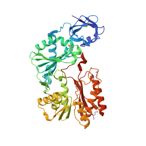
    3C0K

  • PubMed Abstract: 

    Methylation is the most common RNA modification in the three domains of life. Transfer of the methyl group from S-adenosyl-l-methionine (AdoMet) to specific atoms of RNA nucleotides is catalyzed by methyltransferase (MTase) enzymes. The rRNA MTase RlmI (rRNA large subunit methyltransferase gene I; previously known as YccW) specifically modifies Escherichia coli 23S rRNA at nucleotide C1962 to form 5-methylcytosine. Here, we report the crystal structure of RlmI refined at 2 A to a final R-factor of 0.194 (R(free)=0.242). The RlmI molecule comprises three domains: the N-terminal PUA domain; the central domain, which resembles a domain previously found in RNA:5-methyluridine MTases; and the C-terminal catalytic domain, which contains the AdoMet-binding site. The central and C-terminal domains are linked by a beta-hairpin structure that has previously been observed in several MTases acting on nucleic acids or proteins. Based on bioinformatics analyses, we propose a model for the RlmI-AdoMet-RNA complex. Comparative structural analyses of RlmI and its homologs provide insight into the potential function of several structures that have been solved by structural genomics groups and furthermore indicate that the evolutionary paths of RNA and DNA 5-methyluridine and 5-methylcytosine MTases have been closely intertwined.


  • Organizational Affiliation
    • Department of Biological Sciences, National University of Singapore, Singapore.

Macromolecule Content 

  • Total Structure Weight: 89.76 kDa 
  • Atom Count: 6,587 
  • Modeled Residue Count: 790 
  • Deposited Residue Count: 792 
  • Unique protein chains: 1

Macromolecules

Find similar proteins by:|  3D Structure
Entity ID: 1
MoleculeChains  Sequence LengthOrganismDetailsImage
UPF0064 protein yccW
A, B
396Escherichia coli K-12Mutation(s): 0 
EC: 2.1.1 (PDB Primary Data), 2.1.1.191 (UniProt)
UniProt
Find proteins for P75876 (Escherichia coli (strain K12))
Explore P75876 
Go to UniProtKB:  P75876
Entity Groups
Sequence Clusters30% Identity50% Identity70% Identity90% Identity95% Identity100% Identity
UniProt GroupP75876
Sequence Annotations
Expand
Reference Sequence

Small Molecules

Modified Residues  1 Unique
IDChains TypeFormula2D DiagramParent
MSE
Query on MSE
A, B
L-PEPTIDE LINKINGC5 H11 N O2 SeMET

Experimental Data & Validation

Experimental Data

  • Method: X-RAY DIFFRACTION
  • Resolution: 2.00 Å
  • R-Value Free:  0.242 (Depositor), 0.199 (DCC) 
  • R-Value Work:  0.194 (Depositor), 0.203 (DCC) 
Space Group: P 1 21 1
Unit Cell:
Length ( Å )Angle ( ˚ )
a = 64.312α = 90
b = 80.875β = 98.91
c = 66.155γ = 90
Software Package:
Software NamePurpose
CBASSdata collection
SnBphasing
CNSrefinement
HKL-2000data reduction
HKL-2000data scaling

Structure Validation

View Full Validation Report


Entry History 

Deposition Data

Revision History  (Full details and data files)

  • Version 1.0: 2008-11-04
    Type: Initial release
  • Version 1.1: 2011-07-13
    Changes: Version format compliance
  • Version 1.2: 2024-10-30
    Changes: Data collection, Database references, Derived calculations, Structure summary