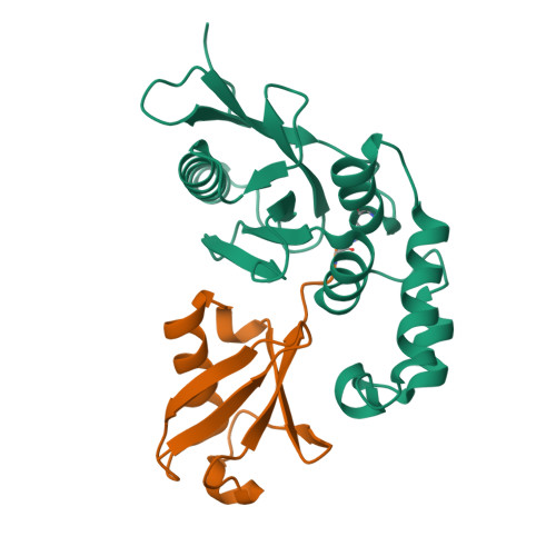
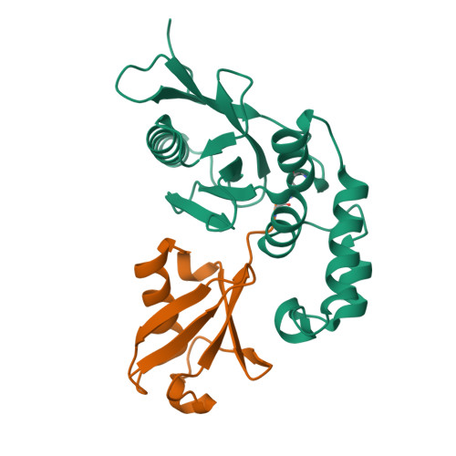
3BY4

Structure of Ovarian Tumor (OTU) domain in complex with Ubiquitin


Experimental Data Snapshot

  • Method: X-RAY DIFFRACTION
  • Resolution: 1.55 Å
  • R-Value Free: 0.211 
  • R-Value Work: 0.189 
  • R-Value Observed: 0.191 

wwPDB Validation   3D Report Full Report


This is version 1.2 of the entry. See complete history


Literature

Structural basis for ubiquitin recognition by the Otu1 ovarian tumor domain protein

Messick, T.E.Russell, N.S.Iwata, A.J.Sarachan, K.L.Shiekhattar, R.Shanks, J.R.Reyes-Turcu, F.E.Wilkinson, K.D.Marmorstein, R.

(2008) J Biol Chem 283: 11038-11049

  • DOI: https://doi.org/10.1074/jbc.M704398200
  • Primary Citation of Related Structures:  
    3BY4, 3C0R

  • PubMed Abstract: 

    Ubiquitination of proteins modifies protein function by either altering their activities, promoting their degradation, or altering their subcellular localization. Deubiquitinating enzymes are proteases that reverse this ubiquitination. Previous studies demonstrate that proteins that contain an ovarian tumor (OTU) domain possess deubiquitinating activity. This domain of approximately 130 amino acids is weakly similar to the papain family of proteases and is highly conserved from yeast to mammals. Here we report structural and functional studies on the OTU domain-containing protein from yeast, Otu1. We show that Otu1 binds polyubiquitin chain analogs more tightly than monoubiquitin and preferentially hydrolyzes longer polyubiquitin chains with Lys(48) linkages, having little or no activity on Lys(63)- and Lys(29)-linked chains. We also show that Otu1 interacts with Cdc48, a regulator of the ER-associated degradation pathway. We also report the x-ray crystal structure of the OTU domain of Otu1 covalently complexed with ubiquitin and carry out structure-guided mutagenesis revealing a novel mode of ubiquitin recognition and a variation on the papain protease catalytic site configuration that appears to be conserved within the OTU family of ubiquitin hydrolases. Together, these studies provide new insights into ubiquitin binding and hydrolysis by yeast Otu1 and other OTU domain-containing proteins.


  • Organizational Affiliation

    Wistar Institute, Philadelphia, Pennsylvania 19104, USA.


Macromolecules
Find similar proteins by:  (by identity cutoff)  |  3D Structure
Entity ID: 1
MoleculeChains Sequence LengthOrganismDetailsImage
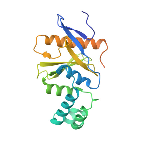
Ubiquitin thioesterase OTU1212Saccharomyces cerevisiaeMutation(s): 0 
Gene Names: Otu1
EC: 3.4.19.12
UniProt
Find proteins for P43558 (Saccharomyces cerevisiae (strain ATCC 204508 / S288c))
Explore P43558 
Go to UniProtKB:  P43558
Entity Groups  
Sequence Clusters30% Identity50% Identity70% Identity90% Identity95% Identity100% Identity
UniProt GroupP43558
Sequence Annotations
Expand
  • Reference Sequence
Find similar proteins by:  (by identity cutoff)  |  3D Structure
Entity ID: 2
MoleculeChains Sequence LengthOrganismDetailsImage
Ubiquitin75Homo sapiensMutation(s): 0 
Gene Names: ubiquitin B
UniProt & NIH Common Fund Data Resources
Find proteins for P0CG48 (Homo sapiens)
Explore P0CG48 
Go to UniProtKB:  P0CG48
PHAROS:  P0CG48
GTEx:  ENSG00000150991 
Entity Groups  
Sequence Clusters30% Identity50% Identity70% Identity90% Identity95% Identity100% Identity
UniProt GroupP0CG48
Sequence Annotations
Expand
  • Reference Sequence
Small Molecules
Ligands 1 Unique
IDChains Name / Formula / InChI Key2D Diagram3D Interactions
3CN
Query on 3CN

Download Ideal Coordinates CCD File 
C [auth B]3-AMINOPROPANE
C3 H9 N
WGYKZJWCGVVSQN-UHFFFAOYSA-N
Experimental Data & Validation

Experimental Data

  • Method: X-RAY DIFFRACTION
  • Resolution: 1.55 Å
  • R-Value Free: 0.211 
  • R-Value Work: 0.189 
  • R-Value Observed: 0.191 
  • Space Group: P 21 21 21
Unit Cell:
Length ( Å )Angle ( ˚ )
a = 46.304α = 90
b = 73.215β = 90
c = 88.88γ = 90
Software Package:
Software NamePurpose
DENZOdata reduction
SCALEPACKdata scaling
SHARPphasing
SOLOMONphasing
PHENIXrefinement
PDB_EXTRACTdata extraction
HKL-2000data collection

Structure Validation

View Full Validation Report


Entry History 

Deposition Data

Revision History  (Full details and data files)

  • Version 1.0: 2008-02-19
    Type: Initial release
  • Version 1.1: 2011-07-13
    Changes: Version format compliance
  • Version 1.2: 2017-10-25
    Changes: Refinement description