3BVS | pdb_00003bvs

Crystal Structure of Bacillus cereus Alkylpurine DNA Glycosylase AlkD


Experimental Data Snapshot

  • Method: X-RAY DIFFRACTION
  • Resolution: 2.10 Å
  • R-Value Free: 
    0.227 (Depositor), 0.233 (DCC) 
  • R-Value Work: 
    0.188 (Depositor), 0.191 (DCC) 
  • R-Value Observed: 
    0.190 (Depositor) 

wwPDB Validation 3D Report Full Report

Validation slider image for 3BVS

This is version 1.2 of the entry. See complete history

Literature

A New Protein Architecture for Processing Alkylation Damaged DNA: The Crystal Structure of DNA Glycosylase AlkD.

Rubinson, E.H.Metz, A.H.O'Quin, J.Eichman, B.F.

(2008) J Mol Biology 381: 13-23

  • DOI: https://doi.org/10.1016/j.jmb.2008.05.078
  • Primary Citation Related Structures: 
    3BVS

  • PubMed Abstract: 

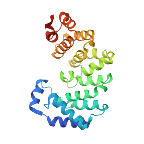
    DNA glycosylases safeguard the genome by locating and excising chemically modified bases from DNA. AlkD is a recently discovered bacterial DNA glycosylase that removes positively charged methylpurines from DNA, and was predicted to adopt a protein fold distinct from from those of other DNA repair proteins. The crystal structure of Bacillus cereus AlkD presented here shows that the protein is composed exclusively of helical HEAT-like repeats, which form a solenoid perfectly shaped to accommodate a DNA duplex on the concave surface. Structural analysis of the variant HEAT repeats in AlkD provides a rationale for how this protein scaffolding motif has been modified to bind DNA. We report 7mG excision and DNA binding activities of AlkD mutants, along with a comparison of alkylpurine DNA glycosylase structures. Together, these data provide important insight into the requirements for alkylation repair within DNA and suggest that AlkD utilizes a novel strategy to manipulate DNA in its search for alkylpurine bases.


  • Organizational Affiliation
    • Department of Biological Sciences and Center for Structural Biology, Vanderbilt University, Nashville, TN 37232, USA.

Macromolecule Content 

  • Total Structure Weight: 28.42 kDa 
  • Atom Count: 1,974 
  • Modeled Residue Count: 227 
  • Deposited Residue Count: 239 
  • Unique protein chains: 1

Macromolecules

Find similar proteins by:|  3D Structure
Entity ID: 1
MoleculeChains  Sequence LengthOrganismDetailsImage
Alkylpurine DNA Glycosylase AlkD239Bacillus cereusMutation(s): 0 
Gene Names: AlkD
UniProt
Find proteins for Q816E8 (Bacillus cereus (strain ATCC 14579 / DSM 31 / CCUG 7414 / JCM 2152 / NBRC 15305 / NCIMB 9373 / NCTC 2599 / NRRL B-3711))
Explore Q816E8 
Go to UniProtKB:  Q816E8
Entity Groups
Sequence Clusters30% Identity50% Identity70% Identity90% Identity95% Identity100% Identity
UniProt GroupQ816E8
Sequence Annotations
Expand
Reference Sequence

Experimental Data & Validation

Experimental Data

  • Method: X-RAY DIFFRACTION
  • Resolution: 2.10 Å
  • R-Value Free:  0.227 (Depositor), 0.233 (DCC) 
  • R-Value Work:  0.188 (Depositor), 0.191 (DCC) 
  • R-Value Observed: 0.190 (Depositor) 
Space Group: P 43
Unit Cell:
Length ( Å )Angle ( ˚ )
a = 77.917α = 90
b = 77.917β = 90
c = 55.102γ = 90
Software Package:
Software NamePurpose
REFMACrefinement
MAR345dtbdata collection
HKL-2000data reduction
HKL-2000data scaling
SHARPphasing

Structure Validation

View Full Validation Report


Entry History 

Deposition Data

Revision History  (Full details and data files)

  • Version 1.0: 2008-07-15
    Type: Initial release
  • Version 1.1: 2011-07-13
    Changes: Advisory, Version format compliance
  • Version 1.2: 2024-02-21
    Changes: Data collection, Database references