Crystal Structure of Human Liver {Delta}4-3-Ketosteroid 5{beta}-Reductase (AKR1D1) and Implications for Substrate Binding and Catalysis.
Di Costanzo, L., Drury, J.E., Penning, T.M., Christianson, D.W.(2008) J Biological Chem 283: 16830-16839
- PubMed: 18407998 Search on PubMedSearch on PubMed Central
- DOI: https://doi.org/10.1074/jbc.M801778200
- Primary Citation Related Structures:
3BUR, 3BUV, 3BV7, 3CMF, 3COT - PubMed Abstract:
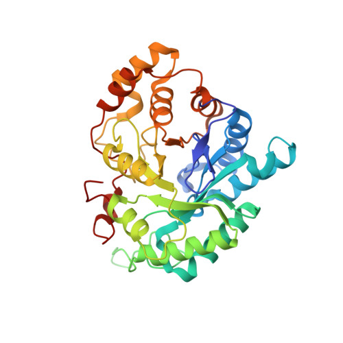
AKR1D1 (steroid 5beta-reductase) reduces all Delta(4)-3-ketosteroids to form 5beta-dihydrosteroids, a first step in the clearance of steroid hormones and an essential step in the synthesis of all bile acids. The reduction of the carbon-carbon double bond in an alpha,beta-unsaturated ketone by 5beta-reductase is a unique reaction in steroid enzymology because hydride transfer from NADPH to the beta-face of a Delta(4)-3-ketosteroid yields a cis-A/B-ring configuration with an approximately 90 degrees bend in steroid structure. Here, we report the first x-ray crystal structure of a mammalian steroid hormone carbon-carbon double bond reductase, human Delta(4)-3-ketosteroid 5beta-reductase (AKR1D1), and its complexes with intact substrates. We have determined the structures of AKR1D1 complexes with NADP(+) at 1.79- and 1.35-A resolution (HEPES bound in the active site), NADP(+) and cortisone at 1.90-A resolution, NADP(+) and progesterone at 2.03-A resolution, and NADP(+) and testosterone at 1.62-A resolution. Complexes with cortisone and progesterone reveal productive substrate binding orientations based on the proximity of each steroid carbon-carbon double bond to the re-face of the nicotinamide ring of NADP(+). This orientation would permit 4-pro-(R)-hydride transfer from NADPH. Each steroid carbonyl accepts hydrogen bonds from catalytic residues Tyr(58) and Glu(120). The Y58F and E120A mutants are devoid of activity, supporting a role for this dyad in the catalytic mechanism. Intriguingly, testosterone binds nonproductively, thereby rationalizing the substrate inhibition observed with this particular steroid. The locations of disease-linked mutations thought to be responsible for bile acid deficiency are also revealed.
- Roy and Diana Vagelos Laboratories, Department of Chemistry, University of Pennsylvania, Philadelphia, Pennsylvania 19104-6323, USA.
Organizational Affiliation: