3BS9 | pdb_00003bs9

X-ray structure of human TIA-1 RRM2


Experimental Data Snapshot

  • Method: X-RAY DIFFRACTION
  • Resolution: 1.95 Å
  • R-Value Free: 
    0.245 (Depositor), 0.241 (DCC) 
  • R-Value Work: 
    0.216 (Depositor), 0.211 (DCC) 

Starting Model: experimental
View more details

wwPDB Validation 3D Report Full Report

Validation slider image for 3BS9

This is version 1.3 of the entry. See complete history

Literature

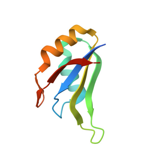
Structure of the central RNA recognition motif of human TIA-1 at 1.95A resolution.

Kumar, A.O.Swenson, M.C.Benning, M.M.Kielkopf, C.L.

(2008) Biochem Biophys Res Commun 367: 813-819

  • DOI: https://doi.org/10.1016/j.bbrc.2008.01.027
  • Primary Citation Related Structures: 
    3BS9

  • PubMed Abstract: 

    T-cell-restricted intracellular antigen-1 (TIA-1) regulates alternative pre-mRNA splicing in the nucleus, and mRNA translation in the cytoplasm, by recognizing uridine-rich sequences of RNAs. As a step towards understanding RNA recognition by this regulatory factor, the X-ray structure of the central RNA recognition motif (RRM2) of human TIA-1 is presented at 1.95A resolution. Comparison with structurally homologous RRM-RNA complexes identifies residues at the RNA interfaces that are conserved in TIA-1-RRM2. The versatile capability of RNP motifs to interact with either proteins or RNA is reinforced by symmetry-related protein-protein interactions mediated by the RNP motifs of TIA-1-RRM2. Importantly, the TIA-1-RRM2 structure reveals the locations of mutations responsible for inhibiting nuclear import. In contrast with previous assumptions, the mutated residues are buried within the hydrophobic interior of the domain, where they would be likely to destabilize the RRM fold rather than directly inhibit RNA binding.


  • Organizational Affiliation
    • Department of Biochemistry and Biophysics, University of Rochester School of Medicine and Dentistry, Rochester, NY 14642, USA.

Macromolecule Content 

  • Total Structure Weight: 19.8 kDa 
  • Atom Count: 1,344 
  • Modeled Residue Count: 158 
  • Deposited Residue Count: 174 
  • Unique protein chains: 1

Macromolecules

Find similar proteins by:|  3D Structure
Entity ID: 1
MoleculeChains  Sequence LengthOrganismDetailsImage
Nucleolysin TIA-1 isoform p40
A, B
87Homo sapiensMutation(s): 3 
Gene Names: TIA1
UniProt & NIH Common Fund Data Resources
Find proteins for P31483 (Homo sapiens)
Explore P31483 
Go to UniProtKB:  P31483
PHAROS:  P31483
GTEx:  ENSG00000116001 
Entity Groups
Sequence Clusters30% Identity50% Identity70% Identity90% Identity95% Identity100% Identity
UniProt GroupP31483
Sequence Annotations
Expand
Reference Sequence

Experimental Data & Validation

Experimental Data

  • Method: X-RAY DIFFRACTION
  • Resolution: 1.95 Å
  • R-Value Free:  0.245 (Depositor), 0.241 (DCC) 
  • R-Value Work:  0.216 (Depositor), 0.211 (DCC) 
Space Group: P 61
Unit Cell:
Length ( Å )Angle ( ˚ )
a = 56.5α = 90
b = 56.5β = 90
c = 76.7γ = 120
Software Package:
Software NamePurpose
PROTEUM PLUSdata collection
PHASERphasing
CNSrefinement
PROTEUM PLUSdata reduction
PROTEUM PLUSdata scaling

Structure Validation

View Full Validation Report


Entry History 

Deposition Data

Revision History  (Full details and data files)

  • Version 1.0: 2008-01-15
    Type: Initial release
  • Version 1.1: 2011-07-13
    Changes: Version format compliance
  • Version 1.2: 2021-10-20
    Changes: Database references, Derived calculations
  • Version 1.3: 2023-08-30
    Changes: Data collection, Refinement description