3BQU

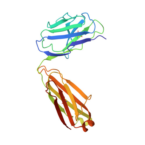
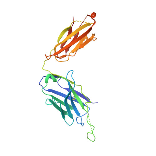
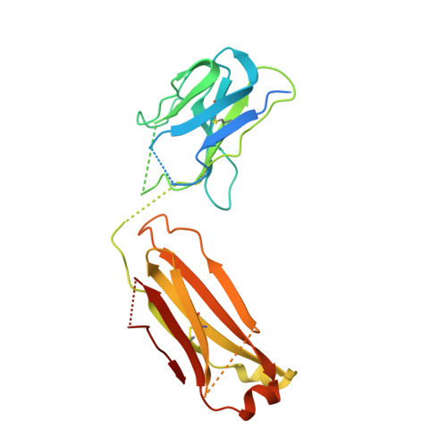
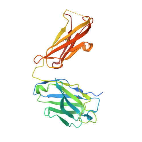
Crystal Structure of the 2F5 Fab'-3H6 Fab Complex


Experimental Data Snapshot

  • Method: X-RAY DIFFRACTION
  • Resolution: 3.00 Å
  • R-Value Free: 0.295 
  • R-Value Work: 0.276 
  • R-Value Observed: 0.276 

Starting Models: experimental
View more details

wwPDB Validation   3D Report Full Report


This is version 1.2 of the entry. See complete history


Literature

Crystal structure of the complex between the F(ab)' fragment of the cross-neutralizing anti-HIV-1 antibody 2F5 and the F(ab) fragment of its anti-idiotypic antibody 3H6.

Bryson, S.Julien, J.P.Isenman, D.E.Kunert, R.Katinger, H.Pai, E.F.

(2008) J Mol Biol 382: 910-919

  • DOI: https://doi.org/10.1016/j.jmb.2008.07.057
  • Primary Citation of Related Structures:  
    3BQU

  • PubMed Abstract: 

    The monoclonal antibody 2F5 neutralizes a broad range of human immunodeficiency virus-1 isolates via a conserved epitope on the viral glycoprotein gp41. The conformation of the principal epitope is a type I beta-turn centered on gp41 residues (664)DKW(666); in addition, binding studies indicate that residues N- and C-terminal to this core as well as structurally more distant parts of gp41 also contribute to the interaction. Ab2/3H6 is an anti-idiotypic antibody that inhibits the interaction between 2F5 and gp41 and as such, Ab2/3H6 may, in principle, possess a paratope that mimics the gp41 epitope. To establish the potential of Ab2/3H6 to serve as a guide for the design of vaccine components against human immunodeficiency virus, we investigated the crystal structure of the heterodimeric complex of Ab2/3H6 F(ab) and 2F5 F(ab)'. Ab2/3H6 F(ab) binds to 2F5 F(ab)' via a helix-like protrusion formed by residues (58(H))RYSPSLNTRL(67(H)) of the 2F5 F(ab)' variable domain and proximal to but not overlapping with the gp41 (664)DKW(666) epitope-binding pocket. This defines Ab2/3H6 as an anti-idiotypic antibody of the Ab2gamma class, i.e., an antigen-inhibitable idiotype that does not carry the internal image of the linear primary gp41 (662)ELDKWAS(668) epitope.


  • Organizational Affiliation

    Department of Biochemistry, University of Toronto, 1 King's College Circle, Toronto, Ontario, Canada M5S 1A8.


Macromolecules
Find similar proteins by:  (by identity cutoff)  |  3D Structure
Entity ID: 1
MoleculeChains Sequence LengthOrganismDetailsImage
2F5 Fab' light chain214Homo sapiensMutation(s): 0 
Entity Groups  
Sequence Clusters30% Identity50% Identity70% Identity90% Identity95% Identity100% Identity
Sequence Annotations
Expand
  • Reference Sequence
Find similar proteins by:  (by identity cutoff)  |  3D Structure
Entity ID: 2
MoleculeChains Sequence LengthOrganismDetailsImage
2F5 Fab' heavy chain235Homo sapiensMutation(s): 0 
UniProt
Find proteins for Q6PYX1 (Homo sapiens)
Explore Q6PYX1 
Go to UniProtKB:  Q6PYX1
Entity Groups  
Sequence Clusters30% Identity50% Identity70% Identity90% Identity95% Identity100% Identity
UniProt GroupQ6PYX1
Sequence Annotations
Expand
  • Reference Sequence
Find similar proteins by:  (by identity cutoff)  |  3D Structure
Entity ID: 3
MoleculeChains Sequence LengthOrganismDetailsImage
3H6 Fab light chain233Mus musculusMutation(s): 0 
UniProt
Find proteins for A2NHM3 (Mus musculus)
Explore A2NHM3 
Go to UniProtKB:  A2NHM3
Entity Groups  
Sequence Clusters30% Identity50% Identity70% Identity90% Identity95% Identity100% Identity
UniProt GroupA2NHM3
Sequence Annotations
Expand
  • Reference Sequence
Find similar proteins by:  (by identity cutoff)  |  3D Structure
Entity ID: 4
MoleculeChains Sequence LengthOrganismDetailsImage
3H6 Fab heavy chain241Mus musculusMutation(s): 0 
UniProt
Find proteins for P01756 (Mus musculus)
Explore P01756 
Go to UniProtKB:  P01756
Entity Groups  
Sequence Clusters30% Identity50% Identity70% Identity90% Identity95% Identity100% Identity
UniProt GroupP01756
Sequence Annotations
Expand
  • Reference Sequence
Small Molecules
Ligands 1 Unique
IDChains Name / Formula / InChI Key2D Diagram3D Interactions
SO4
Query on SO4

Download Ideal Coordinates CCD File 
E [auth D]SULFATE ION
O4 S
QAOWNCQODCNURD-UHFFFAOYSA-L
Experimental Data & Validation

Experimental Data

  • Method: X-RAY DIFFRACTION
  • Resolution: 3.00 Å
  • R-Value Free: 0.295 
  • R-Value Work: 0.276 
  • R-Value Observed: 0.276 
  • Space Group: P 21 21 21
Unit Cell:
Length ( Å )Angle ( ˚ )
a = 67.44α = 90
b = 98.33β = 90
c = 154.39γ = 90
Software Package:
Software NamePurpose
MAR345data collection
CNSrefinement
DENZOdata reduction
SCALEPACKdata scaling
CNSphasing

Structure Validation

View Full Validation Report


Entry History 

Deposition Data

Revision History  (Full details and data files)

  • Version 1.0: 2008-10-21
    Type: Initial release
  • Version 1.1: 2011-07-13
    Changes: Version format compliance
  • Version 1.2: 2023-08-30
    Changes: Data collection, Database references, Derived calculations, Refinement description