3BOG | pdb_00003bog

Snow Flea Antifreeze Protein Quasi-racemate


Experimental Data Snapshot

  • Method: X-RAY DIFFRACTION
  • Resolution: 1.20 Å
  • R-Value Free: 
    0.192 (Depositor), 0.193 (DCC) 
  • R-Value Work: 
    0.160 (Depositor), 0.160 (DCC) 
  • R-Value Observed: 
    0.162 (Depositor) 

wwPDB Validation 3D Report Full Report

Validation slider image for 3BOG

This is version 2.2 of the entry. See complete history

Literature

X-ray structure of snow flea antifreeze protein determined by racemic crystallization of synthetic protein enantiomers

Pentelute, B.L.Gates, Z.P.Tereshko, V.Dashnau, J.L.Vanderkooi, J.M.Kossiakoff, A.A.Kent, S.B.

(2008) J Am Chem Soc 130: 9695-9701

  • DOI: https://doi.org/10.1021/ja8013538
  • Primary Citation Related Structures: 
    2PNE, 3BOG, 3BOI

  • PubMed Abstract: 

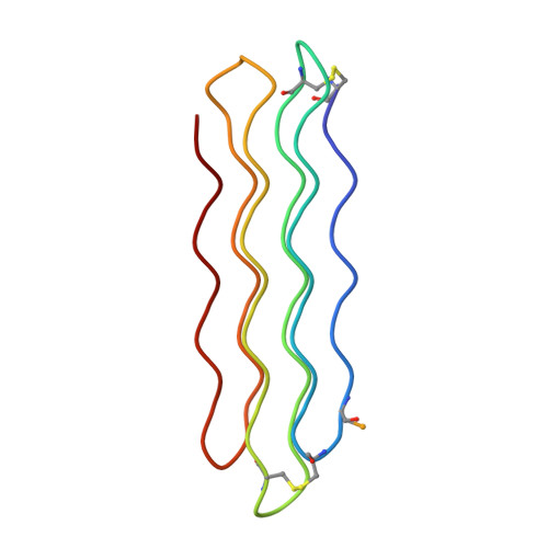
    Chemical protein synthesis and racemic protein crystallization were used to determine the X-ray structure of the snow flea antifreeze protein (sfAFP). Crystal formation from a racemic solution containing equal amounts of the chemically synthesized proteins d-sfAFP and l-sfAFP occurred much more readily than for l-sfAFP alone. More facile crystal formation also occurred from a quasi-racemic mixture of d-sfAFP and l-Se-sfAFP, a chemical protein analogue that contains an additional -SeCH2- moiety at one residue and thus differs slightly from the true enantiomer. Multiple wavelength anomalous dispersion (MAD) phasing from quasi-racemate crystals was then used to determine the X-ray structure of the sfAFP protein molecule. The resulting model was used to solve by molecular replacement the X-ray structure of l-sfAFP to a resolution of 0.98 A. The l-sfAFP molecule is made up of six antiparallel left-handed PPII helixes, stacked in two sets of three, to form a compact brick-like structure with one hydrophilic face and one hydrophobic face. This is a novel experimental protein structure and closely resembles a structural model proposed for sfAFP. These results illustrate the utility of total chemical synthesis combined with racemic crystallization and X-ray crystallography for determining the unknown structure of a protein.


  • Organizational Affiliation
    • Institute for Biophysical Dynamics, University of Chicago, Chicago, Illinois 60637, USA.

Macromolecule Content 

  • Total Structure Weight: 26.14 kDa 
  • Atom Count: 2,303 
  • Modeled Residue Count: 324 
  • Deposited Residue Count: 324 
  • Unique protein chains: 2

Macromolecules

Find similar proteins by:|  3D Structure
Entity ID: 1
MoleculeChains  Sequence LengthOrganismDetailsImage
6.5 kDa glycine-rich antifreeze protein
A, B
81Hypogastrura harveyiMutation(s): 1 
UniProt
Find proteins for Q38PT6 (Hypogastrura harveyi)
Explore Q38PT6 
Go to UniProtKB:  Q38PT6
Entity Groups
Sequence Clusters30% Identity50% Identity70% Identity90% Identity95% Identity100% Identity
UniProt GroupQ38PT6
Sequence Annotations
Expand
Reference Sequence
Find similar proteins by:|  3D Structure
Entity ID: 2
MoleculeChains  Sequence LengthOrganismDetailsImage
6.5 kDa glycine-rich antifreeze protein
C, D
81Hypogastrura harveyiMutation(s): 0 
UniProt
Find proteins for Q38PT6 (Hypogastrura harveyi)
Explore Q38PT6 
Go to UniProtKB:  Q38PT6
Entity Groups
Sequence Clusters30% Identity50% Identity70% Identity90% Identity95% Identity100% Identity
UniProt GroupQ38PT6
Sequence Annotations
Expand
Reference Sequence

Small Molecules

Ligands 1 Unique
IDChains Name / Formula / InChI Key2D Diagram3D Interactions
UNL

Query on UNL


Download:Ideal Coordinates CCD File
E [auth B]Unknown ligand
Modified Residues  1 Unique
IDChains TypeFormula2D DiagramParent
SYS
Query on SYS
A, B
L-PEPTIDE LINKINGC5 H10 N2 O3 SeSEC
DAL
Query on DAL
C, D
D-PEPTIDE LINKINGC3 H7 N O2

--

DAR
Query on DAR
C, D
D-PEPTIDE LINKINGC6 H15 N4 O2

--

DAS
Query on DAS
C, D
D-PEPTIDE LINKINGC4 H7 N O4

--

DCY
Query on DCY
C, D
D-PEPTIDE LINKINGC3 H7 N O2 S

--

DHI
Query on DHI
C, D
D-PEPTIDE LINKINGC6 H10 N3 O2

--

DLY
Query on DLY
C, D
D-PEPTIDE LINKINGC6 H14 N2 O2

--

DPR
Query on DPR
C, D
D-PEPTIDE LINKINGC5 H9 N O2

--

DSG
Query on DSG
C, D
D-PEPTIDE LINKINGC4 H8 N2 O3

--

DSN
Query on DSN
C, D
D-PEPTIDE LINKINGC3 H7 N O3

--

DTH
Query on DTH
C, D
D-PEPTIDE LINKINGC4 H9 N O3

--

DVA
Query on DVA
C, D
D-PEPTIDE LINKINGC5 H11 N O2

--

Experimental Data & Validation

Experimental Data

  • Method: X-RAY DIFFRACTION
  • Resolution: 1.20 Å
  • R-Value Free:  0.192 (Depositor), 0.193 (DCC) 
  • R-Value Work:  0.160 (Depositor), 0.160 (DCC) 
  • R-Value Observed: 0.162 (Depositor) 
Space Group: P 1
Unit Cell:
Length ( Å )Angle ( ˚ )
a = 28.64α = 88.62
b = 32.359β = 89.31
c = 59.699γ = 72.84
Software Package:
Software NamePurpose
REFMACrefinement
HKL-2000data reduction
HKL-2000data scaling
SHELXDphasing
SOLVEphasing
RESOLVEphasing

Structure Validation

View Full Validation Report


Entry History 

Deposition Data

Revision History  (Full details and data files)

  • Version 1.0: 2008-09-23
    Type: Initial release
  • Version 1.1: 2011-07-13
    Changes: Version format compliance
  • Version 2.0: 2018-01-24
    Changes: Polymer sequence, Source and taxonomy, Structure summary
  • Version 2.1: 2019-08-21
    Changes: Advisory, Data collection, Derived calculations
  • Version 2.2: 2021-10-20
    Changes: Database references, Derived calculations