3BIA | pdb_00003bia

Tim-4 in complex with sodium potassium tartrate


Experimental Data Snapshot

  • Method: X-RAY DIFFRACTION
  • Resolution: 2.20 Å
  • R-Value Free: 
    0.238 (Depositor), 0.239 (DCC) 
  • R-Value Work: 
    0.218 (Depositor), 0.218 (DCC) 
  • R-Value Observed: 
    0.219 (Depositor) 

Starting Model: experimental
View more details

wwPDB Validation 3D Report Full Report

Validation slider image for 3BIA

This is version 1.3 of the entry. See complete history

Literature

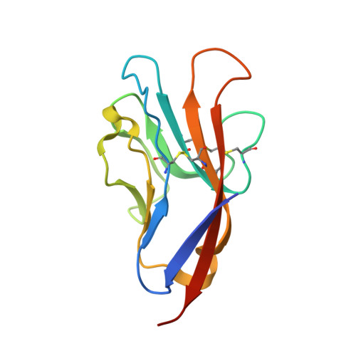
Structures of T Cell Immunoglobulin Mucin Protein 4 Show a Metal-Ion-Dependent Ligand Binding Site where Phosphatidylserine Binds.

Santiago, C.Ballesteros, A.Martinez-Munoz, L.Mellado, M.Kaplan, G.G.Freeman, G.J.Casasnovas, J.M.

(2007) Immunity 27: 941-951

  • DOI: https://doi.org/10.1016/j.immuni.2007.11.008
  • Primary Citation Related Structures: 
    3BI9, 3BIA, 3BIB

  • PubMed Abstract: 

    The T cell immunoglobulin and mucin domain (TIM) proteins are important regulators of T cell responses. Crystal structures of the murine TIM-4 identified a metal-ion-dependent ligand binding site (MILIBS) in the immunoglobulin (Ig) domain of the TIM family. The characteristic CC' loop of the TIM domain and the hydrophobic FG loop shaped a narrow cavity where acidic compounds penetrate and coordinate to a metal ion bound to conserved residues in the TIM proteins. The structure of phosphatidylserine bound to the Ig domain showed that the hydrophilic head penetrates into the MILIBS and coordinates with the metal ion, whereas the aromatic residues on the tip of the FG loop interacted with the fatty acid chains and could insert into the lipid bilayer. Our results also revealed an important role of the MILIBS in the trafficking of TIM-1 to the cell surface.


  • Organizational Affiliation
    • Centro Nacional de Biotecnología, CSIC, Campus Universidad Autónoma, 28049 Madrid, Spain.

Macromolecule Content 

  • Total Structure Weight: 13.23 kDa 
  • Atom Count: 955 
  • Modeled Residue Count: 110 
  • Deposited Residue Count: 116 
  • Unique protein chains: 1

Macromolecules

Find similar proteins by:|  3D Structure
Entity ID: 1
MoleculeChains  Sequence LengthOrganismDetailsImage
T-cell immunoglobulin and mucin domain-containing protein 4A [auth X]116Mus musculusMutation(s): 0 
Gene Names: Timd4Tim4
UniProt
Find proteins for Q6U7R4 (Mus musculus)
Explore Q6U7R4 
Go to UniProtKB:  Q6U7R4
Entity Groups
Sequence Clusters30% Identity50% Identity70% Identity90% Identity95% Identity100% Identity
UniProt GroupQ6U7R4
Sequence Annotations
Expand
Reference Sequence

Experimental Data & Validation

Experimental Data

  • Method: X-RAY DIFFRACTION
  • Resolution: 2.20 Å
  • R-Value Free:  0.238 (Depositor), 0.239 (DCC) 
  • R-Value Work:  0.218 (Depositor), 0.218 (DCC) 
  • R-Value Observed: 0.219 (Depositor) 
Space Group: P 63 2 2
Unit Cell:
Length ( Å )Angle ( ˚ )
a = 67.77α = 90
b = 67.77β = 90
c = 129.09γ = 120
Software Package:
Software NamePurpose
REFMACrefinement
XDSdata reduction
SCALAdata scaling
PHASERphasing

Structure Validation

View Full Validation Report


Entry History 

Deposition Data

Revision History  (Full details and data files)

  • Version 1.0: 2008-01-01
    Type: Initial release
  • Version 1.1: 2011-07-13
    Changes: Version format compliance
  • Version 1.2: 2024-04-03
    Changes: Data collection, Database references, Derived calculations, Refinement description
  • Version 1.3: 2024-10-16
    Changes: Structure summary