3BCZ | pdb_00003bcz

Crystal structure of Memo


Experimental Data Snapshot

  • Method: X-RAY DIFFRACTION
  • Resolution: 2.10 Å
  • R-Value Free: 
    0.230 (Depositor), 0.186 (DCC) 
  • R-Value Work: 
    0.183 (Depositor), 0.187 (DCC) 
  • R-Value Observed: 
    0.185 (Depositor) 

wwPDB Validation 3D Report Full Report

Validation slider image for 3BCZ

This is version 1.3 of the entry. See complete history

Literature

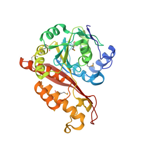
Memo Is Homologous to Nonheme Iron Dioxygenases and Binds an ErbB2-derived Phosphopeptide in Its Vestigial Active Site.

Qiu, C.Lienhard, S.Hynes, N.E.Badache, A.Leahy, D.J.

(2008) J Biological Chem 283: 2734-2740

  • DOI: https://doi.org/10.1074/jbc.M703523200
  • Primary Citation Related Structures: 
    3BCZ, 3BD0

  • PubMed Abstract: 

    Memo (mediator of ErbB2-driven cell motility) is a 297-amino-acid protein recently shown to co-precipitate with the C terminus of ErbB2 and be required for ErbB2-driven cell motility. Memo is not homologous to any known signaling proteins, and how it mediates ErbB2 signals is not known. To provide a molecular basis for understanding Memo function, we have determined and report here the 2.1A crystal structure of human Memo and show it be homologous to class III nonheme iron-dependent dioxygenases, a structural class that now includes a zinc-binding protein of unknown function. No metal binding or enzymatic activity can be detected for Memo, but Memo does bind directly to a specific ErbB2-derived phosphopeptide encompassing Tyr-1227 using its vestigial enzymatic active site. Memo thus represents a new class of phosphotyrosine-binding protein.


  • Organizational Affiliation
    • Department of Biophysics and Biophysical Chemistry, The Johns Hopkins University School of Medicine, 725 N. Wolfe Street, Baltimore, MD 21205, USA.

Macromolecule Content 

  • Total Structure Weight: 135.1 kDa 
  • Atom Count: 9,863 
  • Modeled Residue Count: 1,171 
  • Deposited Residue Count: 1,172 
  • Unique protein chains: 1

Macromolecules

Find similar proteins by:|  3D Structure
Entity ID: 1
MoleculeChains  Sequence LengthOrganismDetailsImage
Protein MEMO1
A, B, C, D
293Homo sapiensMutation(s): 0 
Gene Names: MEMO1C2orf4NS5ATP7
UniProt & NIH Common Fund Data Resources
Find proteins for Q9Y316 (Homo sapiens)
Explore Q9Y316 
Go to UniProtKB:  Q9Y316
PHAROS:  Q9Y316
GTEx:  ENSG00000162959 
Entity Groups
Sequence Clusters30% Identity50% Identity70% Identity90% Identity95% Identity100% Identity
UniProt GroupQ9Y316
Sequence Annotations
Expand
Reference Sequence

Experimental Data & Validation

Experimental Data

  • Method: X-RAY DIFFRACTION
  • Resolution: 2.10 Å
  • R-Value Free:  0.230 (Depositor), 0.186 (DCC) 
  • R-Value Work:  0.183 (Depositor), 0.187 (DCC) 
  • R-Value Observed: 0.185 (Depositor) 
Space Group: P 21 21 2
Unit Cell:
Length ( Å )Angle ( ˚ )
a = 139.826α = 90
b = 88.849β = 90
c = 98.089γ = 90
Software Package:
Software NamePurpose
DENZOdata reduction
SCALEPACKdata scaling
REFMACrefinement
PDB_EXTRACTdata extraction
ADSCdata collection
HKL-2000data reduction
HKL-2000data scaling
SnBphasing

Structure Validation

View Full Validation Report


Entry History 

Deposition Data

  • Released Date: 2007-11-27 
  • Deposition Author(s): Qiu, C.

Revision History  (Full details and data files)

  • Version 1.0: 2007-11-27
    Type: Initial release
  • Version 1.1: 2011-07-13
    Changes: Advisory, Version format compliance
  • Version 1.2: 2017-10-25
    Changes: Refinement description
  • Version 1.3: 2024-11-06
    Changes: Data collection, Database references, Derived calculations, Refinement description, Structure summary