


D-Glucopyranosyl pyrimidine nucleoside binding to muscle glycogen phosphorylase b
Cisma, C., Sovantzis, D.A., Hadjiloi, T., Stathis, D., Gimisis, T., Hayes, J.M., Zographos, S.E., Leonidas, D.D., Chrysina, E.D., Oikonomakos, N.G.To be published.
Experimental Data Snapshot
Starting Model: experimental
View more details
Entity ID: 1 | |||||
|---|---|---|---|---|---|
| Molecule | Chains | Sequence Length | Organism | Details | Image |
| Glycogen phosphorylase, muscle form | 842 | Oryctolagus cuniculus | Mutation(s): 0 EC: 2.4.1.1 | 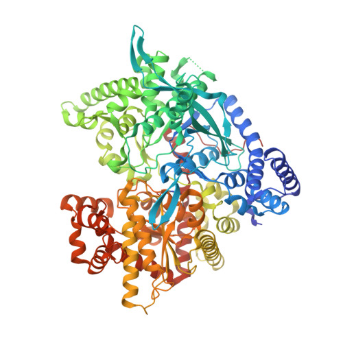 | |
UniProt | |||||
Entity Groups | |||||
| Sequence Clusters | 30% Identity50% Identity70% Identity90% Identity95% Identity100% Identity | ||||
| UniProt Group | P00489 | ||||
Sequence AnnotationsExpand | |||||
Reference Sequence | |||||
| Ligands 1 Unique | |||||
|---|---|---|---|---|---|
| ID | Chains | Name / Formula / InChI Key | 2D Diagram | 3D Interactions | |
| CJB Download:Ideal Coordinates CCD File | B [auth A] | 1-beta-D-glucopyranosylpyrimidine-2,4(1H,3H)-dione C10 H14 N2 O7 PBGBADORVLQASN-DDIGBBAMSA-N |  | ||
| Modified Residues 1 Unique | |||||
|---|---|---|---|---|---|
| ID | Chains | Type | Formula | 2D Diagram | Parent |
| LLP Query on LLP | A | L-PEPTIDE LINKING | C14 H22 N3 O7 P |  | LYS |
| Length ( Å ) | Angle ( ˚ ) |
|---|---|
| a = 128.577 | α = 90 |
| b = 128.577 | β = 90 |
| c = 116.517 | γ = 90 |
| Software Name | Purpose |
|---|---|
| REFMAC | refinement |
| ADSC | data collection |
| DENZO | data reduction |
| SCALEPACK | data scaling |


