3BCG | pdb_00003bcg

Conformational changes of the AcrR regulator reveal a mechanism of induction


Experimental Data Snapshot

  • Method: X-RAY DIFFRACTION
  • Resolution: 2.48 Å
  • R-Value Free: 
    0.268 (Depositor), 0.296 (DCC) 
  • R-Value Work: 
    0.214 (Depositor), 0.305 (DCC) 

wwPDB Validation 3D Report Full Report

Validation slider image for 3BCG

This is version 1.2 of the entry. See complete history

Literature

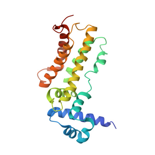
Conformational change of the AcrR regulator reveals a possible mechanism of induction.

Gu, R.Li, M.Su, C.C.Long, F.Routh, M.D.Yang, F.McDermott, G.Yu, E.W.

(2008) Acta Crystallogr Sect F Struct Biol Cryst Commun 64: 584-588

  • DOI: https://doi.org/10.1107/S1744309108016035
  • Primary Citation Related Structures: 
    3BCG

  • PubMed Abstract: 

    The Escherichia coli AcrR multidrug-binding protein represses transcription of acrAB and is induced by many structurally unrelated cytotoxic compounds. The crystal structure of AcrR in space group P222(1) has been reported previously. This P222(1) structure has provided direct information about the multidrug-binding site and important residues for drug recognition. Here, a crystal structure of this regulator in space group P3(1) is presented. Comparison of the two AcrR structures reveals possible mechanisms of ligand binding and AcrR regulation.


  • Organizational Affiliation
    • Department of Physics and Astronomy, Iowa State University, Ames, IA 50011, USA.

Macromolecule Content 

  • Total Structure Weight: 49.6 kDa 
  • Atom Count: 3,428 
  • Modeled Residue Count: 414 
  • Deposited Residue Count: 430 
  • Unique protein chains: 1

Macromolecules

Find similar proteins by:|  3D Structure
Entity ID: 1
MoleculeChains  Sequence LengthOrganismDetailsImage
HTH-type transcriptional regulator acrR
A, B
215Escherichia coli K-12Mutation(s): 0 
Gene Names: acrR
UniProt
Find proteins for P0ACS9 (Escherichia coli (strain K12))
Explore P0ACS9 
Go to UniProtKB:  P0ACS9
Entity Groups
Sequence Clusters30% Identity50% Identity70% Identity90% Identity95% Identity100% Identity
UniProt GroupP0ACS9
Sequence Annotations
Expand
Reference Sequence

Experimental Data & Validation

Experimental Data

  • Method: X-RAY DIFFRACTION
  • Resolution: 2.48 Å
  • R-Value Free:  0.268 (Depositor), 0.296 (DCC) 
  • R-Value Work:  0.214 (Depositor), 0.305 (DCC) 
Space Group: P 31
Unit Cell:
Length ( Å )Angle ( ˚ )
a = 46.66α = 90
b = 46.66β = 90
c = 166.155γ = 120
Software Package:
Software NamePurpose
HKL-2000data collection
SnBphasing
CNSrefinement
HKL-2000data reduction
HKL-2000data scaling

Structure Validation

View Full Validation Report


Entry History 

Deposition Data

Revision History  (Full details and data files)

  • Version 1.0: 2008-02-26
    Type: Initial release
  • Version 1.1: 2011-07-13
    Changes: Version format compliance
  • Version 1.2: 2024-02-21
    Changes: Data collection, Database references