3BCA | pdb_00003bca

Crystal structure of mouse selenocysteine synthase, sodium iodide soak


Experimental Data Snapshot

  • Method: X-RAY DIFFRACTION
  • Resolution: 2.25 Å
  • R-Value Free: 
    0.236 (Depositor), 0.230 (DCC) 
  • R-Value Work: 
    0.183 (Depositor), 0.178 (DCC) 
  • R-Value Observed: 
    0.185 (Depositor) 

wwPDB Validation 3D Report Full Report

Validation slider image for 3BCA

This is version 1.3 of the entry. See complete history

Literature

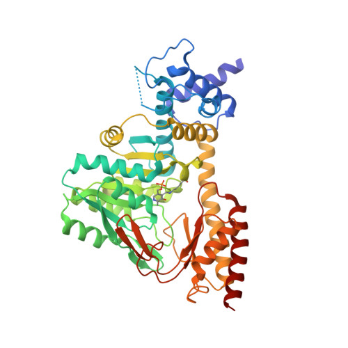
Structure and catalytic mechanism of eukaryotic selenocysteine synthase.

Ganichkin, O.M.Xu, X.M.Carlson, B.A.Mix, H.Hatfield, D.L.Gladyshev, V.N.Wahl, M.C.

(2008) J Biological Chem 283: 5849-5865

  • DOI: https://doi.org/10.1074/jbc.M709342200
  • Primary Citation Related Structures: 
    3BC8, 3BCA, 3BCB

  • PubMed Abstract: 

    In eukaryotes and Archaea, selenocysteine synthase (SecS) converts O-phospho-L-seryl-tRNA [Ser]Sec into selenocysteyl-tRNA [Ser]Sec using selenophosphate as the selenium donor compound. The molecular mechanisms underlying SecS activity are presently unknown. We have delineated a 450-residue core of mouse SecS, which retained full selenocysteyl-tRNA [Ser]Sec synthesis activity, and determined its crystal structure at 1.65 A resolution. SecS exhibits three domains that place it in the fold type I family of pyridoxal phosphate (PLP)-dependent enzymes. Two SecS monomers interact intimately and together build up two identical active sites around PLP in a Schiff-base linkage with lysine 284. Two SecS dimers further associate to form a homotetramer. The N terminus, which mediates tetramer formation, and a large insertion that remodels the active site set SecS aside from other members of the family. The active site insertion contributes to PLP binding and positions a glutamate next to the PLP, where it could repel substrates with a free alpha-carboxyl group, suggesting why SecS does not act on free O-phospho-l-serine. Upon soaking crystals in phosphate buffer, a previously disordered loop within the active site insertion contracted to form a phosphate binding site. Residues that are strictly conserved in SecS orthologs but variant in related enzymes coordinate the phosphate and upon mutation corrupt SecS activity. Modeling suggested that the phosphate loop accommodates the gamma-phosphate moiety of O-phospho-l-seryl-tRNA [Ser]Sec and, after phosphate elimination, binds selenophosphate to initiate attack on the proposed aminoacrylyl-tRNA [Ser]Sec intermediate. Based on these results and on the activity profiles of mechanism-based inhibitors, we offer a detailed reaction mechanism for the enzyme.


  • Organizational Affiliation
    • Max-Planck-Institut für Biophysikalische Chemie, Zelluläre Biochemie/Makromolekulare Röntgenkristallographie, Am Fassberg 11, D-37077 Göttingen, Germany.

Macromolecule Content 

  • Total Structure Weight: 57.85 kDa 
  • Atom Count: 3,778 
  • Modeled Residue Count: 436 
  • Deposited Residue Count: 450 
  • Unique protein chains: 1

Macromolecules

Find similar proteins by:|  3D Structure
Entity ID: 1
MoleculeChains  Sequence LengthOrganismDetailsImage
O-phosphoseryl-tRNA(Sec) selenium transferase450Mus musculusMutation(s): 0 
Gene Names: SepsecsD5Ertd135e
EC: 2.9.1 (PDB Primary Data), 2.9.1.2 (UniProt)
UniProt & NIH Common Fund Data Resources
Find proteins for Q6P6M7 (Mus musculus)
Explore Q6P6M7 
Go to UniProtKB:  Q6P6M7
IMPC:  MGI:1098791
Entity Groups
Sequence Clusters30% Identity50% Identity70% Identity90% Identity95% Identity100% Identity
UniProt GroupQ6P6M7
Sequence Annotations
Expand
Reference Sequence

Small Molecules

Ligands 1 Unique
IDChains Name / Formula / InChI Key2D Diagram3D Interactions
IOD

Query on IOD


Download:Ideal Coordinates CCD File
AA [auth A]
AB [auth A]
B [auth A]
BA [auth A]
BB [auth A]
AA [auth A],
AB [auth A],
B [auth A],
BA [auth A],
BB [auth A],
C [auth A],
CA [auth A],
CB [auth A],
D [auth A],
DA [auth A],
DB [auth A],
E [auth A],
EA [auth A],
EB [auth A],
F [auth A],
FA [auth A],
FB [auth A],
G [auth A],
GA [auth A],
GB [auth A],
H [auth A],
HA [auth A],
HB [auth A],
I [auth A],
IA [auth A],
IB [auth A],
J [auth A],
JA [auth A],
JB [auth A],
K [auth A],
KA [auth A],
KB [auth A],
L [auth A],
LA [auth A],
M [auth A],
MA [auth A],
N [auth A],
NA [auth A],
O [auth A],
OA [auth A],
P [auth A],
PA [auth A],
Q [auth A],
QA [auth A],
R [auth A],
RA [auth A],
S [auth A],
SA [auth A],
T [auth A],
TA [auth A],
U [auth A],
UA [auth A],
V [auth A],
VA [auth A],
W [auth A],
WA [auth A],
X [auth A],
XA [auth A],
Y [auth A],
YA [auth A],
Z [auth A],
ZA [auth A]
IODIDE ION
I
XMBWDFGMSWQBCA-UHFFFAOYSA-M
Modified Residues  1 Unique
IDChains TypeFormula2D DiagramParent
LLP
Query on LLP
A
L-PEPTIDE LINKINGC14 H22 N3 O7 PLYS

Experimental Data & Validation

Experimental Data

  • Method: X-RAY DIFFRACTION
  • Resolution: 2.25 Å
  • R-Value Free:  0.236 (Depositor), 0.230 (DCC) 
  • R-Value Work:  0.183 (Depositor), 0.178 (DCC) 
  • R-Value Observed: 0.185 (Depositor) 
Space Group: I 2 2 2
Unit Cell:
Length ( Å )Angle ( ˚ )
a = 59.222α = 90
b = 138.371β = 90
c = 141.52γ = 90
Software Package:
Software NamePurpose
REFMACrefinement
MAR345dtbdata collection
DENZOdata reduction
SCALEPACKdata scaling
SHELXCDphasing
SHELXEmodel building

Structure Validation

View Full Validation Report


Entry History 

Deposition Data

Revision History  (Full details and data files)

  • Version 1.0: 2007-12-18
    Type: Initial release
  • Version 1.1: 2011-07-13
    Changes: Advisory, Version format compliance
  • Version 1.2: 2017-10-25
    Changes: Refinement description
  • Version 1.3: 2025-03-26
    Changes: Data collection, Database references, Derived calculations, Structure summary