3B9U

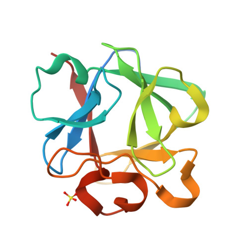
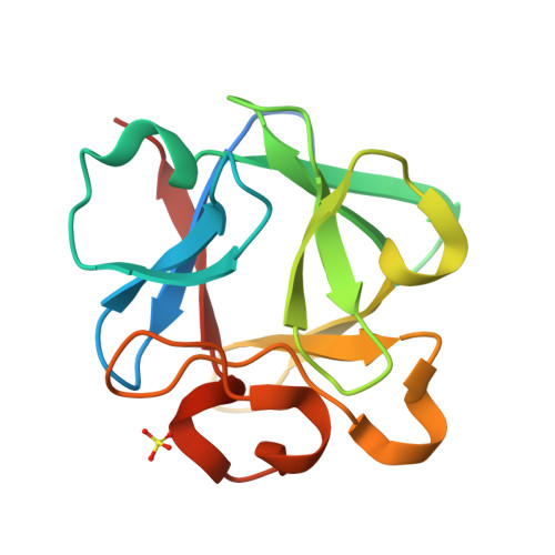
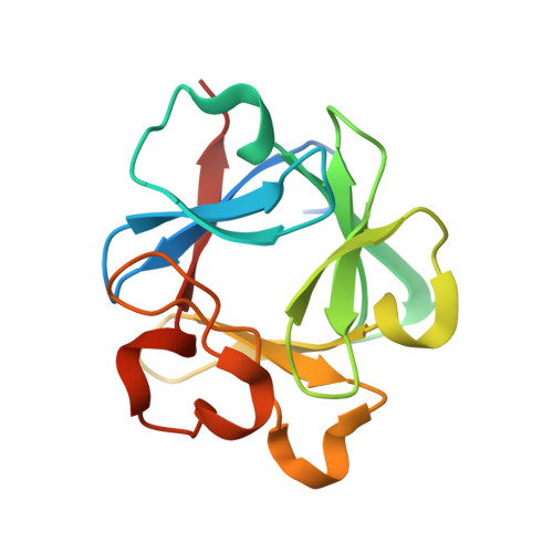
Crystal structure of L26N/D28N/H93G mutant of Human acidic fibroblast growth factor


Experimental Data Snapshot

  • Method: X-RAY DIFFRACTION
  • Resolution: 1.55 Å
  • R-Value Free: 0.200 
  • R-Value Work: 0.191 
  • R-Value Observed: 0.191 

Starting Model: experimental
View more details

wwPDB Validation   3D Report Full Report


This is version 1.3 of the entry. See complete history


Literature

A logical OR redundancy within the Asx-Pro-Asx-Gly type I beta-turn motif.

Lee, J.Dubey, V.K.Longo, L.M.Blaber, M.

(2008) J Mol Biol 377: 1251-1264

  • DOI: https://doi.org/10.1016/j.jmb.2008.01.055
  • Primary Citation of Related Structures:  
    3B9U, 3BA4, 3BA5, 3BA7, 3BAD, 3BAG, 3BAH, 3BAO, 3BAQ, 3BAU, 3BAV, 3BB2

  • PubMed Abstract: 

    Turn secondary structure is essential to the formation of globular protein architecture. Turn structures are, however, much more complex than either alpha-helix or beta-sheet, and the thermodynamics and folding kinetics are poorly understood. Type I beta-turns are the most common type of reverse turn, and they exhibit a statistical consensus sequence of Asx-Pro-Asx-Gly (where Asx is Asp or Asn). A comprehensive series of individual and combined Asx mutations has been constructed within three separate type I 3:5 G1 bulge beta-turns in human fibroblast growth factor-1, and their effects on structure, stability, and folding have been determined. The results show a fundamental logical OR relationship between the Asx residues in the motif, involving H-bond interactions with main-chain amides within the turn. These interactions can be modulated by additional interactions with residues adjacent to the turn at positions i+4 and i+6. The results show that the Asx residues in the turn motif make a substantial contribution to the overall stability of the protein, and the Asx logical OR relationship defines a redundant system that can compensate for deleterious point mutations. The results also show that the stability of the turn is unlikely to be the prime determinant of formation of turn structure in the folding transition state.


  • Organizational Affiliation

    Department of Biomedical Sciences, College of Medicine, Florida State University, Tallahassee, FL 32306-4300, USA.


Macromolecules
Find similar proteins by:  (by identity cutoff)  |  3D Structure
Entity ID: 1
MoleculeChains Sequence LengthOrganismDetailsImage
Heparin-binding growth factor 1146Homo sapiensMutation(s): 3 
Gene Names: FGF1FGFA
UniProt & NIH Common Fund Data Resources
Find proteins for P05230 (Homo sapiens)
Explore P05230 
Go to UniProtKB:  P05230
PHAROS:  P05230
GTEx:  ENSG00000113578 
Entity Groups  
Sequence Clusters30% Identity50% Identity70% Identity90% Identity95% Identity100% Identity
UniProt GroupP05230
Sequence Annotations
Expand
  • Reference Sequence
Small Molecules
Ligands 1 Unique
IDChains Name / Formula / InChI Key2D Diagram3D Interactions
SO4
Query on SO4

Download Ideal Coordinates CCD File 
B [auth A]SULFATE ION
O4 S
QAOWNCQODCNURD-UHFFFAOYSA-L
Experimental Data & Validation

Experimental Data

  • Method: X-RAY DIFFRACTION
  • Resolution: 1.55 Å
  • R-Value Free: 0.200 
  • R-Value Work: 0.191 
  • R-Value Observed: 0.191 
  • Space Group: P 1 21 1
Unit Cell:
Length ( Å )Angle ( ˚ )
a = 33.389α = 90
b = 44.19β = 100.45
c = 47.957γ = 90
Software Package:
Software NamePurpose
CNSrefinement
HKL-2000data collection
HKL-2000data reduction
SCALEPACKdata scaling
CNSphasing

Structure Validation

View Full Validation Report


Entry History 

Deposition Data

Revision History  (Full details and data files)

  • Version 1.0: 2008-04-15
    Type: Initial release
  • Version 1.1: 2011-07-13
    Changes: Version format compliance
  • Version 1.2: 2021-10-20
    Changes: Database references, Derived calculations
  • Version 1.3: 2023-08-30
    Changes: Data collection, Refinement description