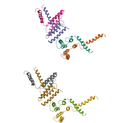
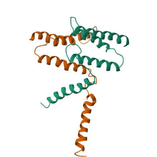
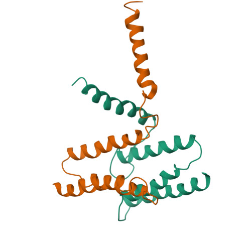
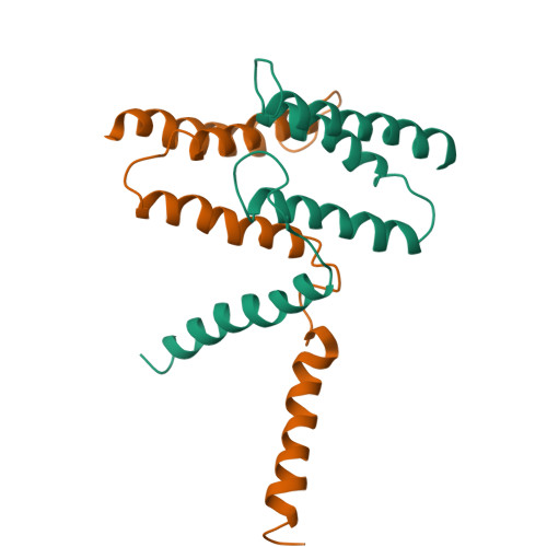
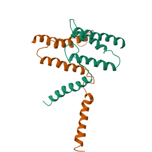
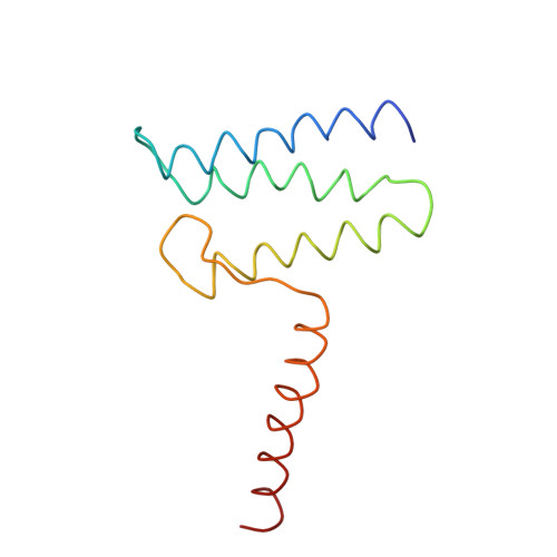
3B61

EmrE multidrug transporter, apo crystal form


Experimental Data Snapshot

  • Method: X-RAY DIFFRACTION
  • Resolution: 4.50 Å
  • R-Value Free: 0.362 
  • R-Value Work: 0.318 

wwPDB Validation   3D Report Full Report


This is version 1.2 of the entry. See complete history


Literature

X-ray structure of EmrE supports dual topology model.

Chen, Y.J.Pornillos, O.Lieu, S.Ma, C.Chen, A.P.Chang, G.

(2007) Proc Natl Acad Sci U S A 104: 18999-19004

  • DOI: https://doi.org/10.1073/pnas.0709387104
  • Primary Citation of Related Structures:  
    3B5D, 3B61, 3B62

  • PubMed Abstract: 

    EmrE, a multidrug transporter from Escherichia coli, functions as a homodimer of a small four-transmembrane protein. The membrane insertion topology of the two monomers is controversial. Although the EmrE protein was reported to have a unique orientation in the membrane, models based on electron microscopy and now defunct x-ray structures, as well as recent biochemical studies, posit an antiparallel dimer. We have now reanalyzed our x-ray data on EmrE. The corrected structures in complex with a transport substrate are highly similar to the electron microscopy structure. The first three transmembrane helices from each monomer surround the substrate binding chamber, whereas the fourth helices participate only in dimer formation. Selenomethionine markers clearly indicate an antiparallel orientation for the monomers, supporting a "dual topology" model.


  • Organizational Affiliation

    Department of Molecular Biology, The Scripps Research Institute, 10550 North Torrey Pines Road, CB-105, La Jolla, CA 92037, USA.


Macromolecules
Find similar proteins by:  (by identity cutoff)  |  3D Structure
Entity ID: 1
MoleculeChains Sequence LengthOrganismDetailsImage
Multidrug transporter emrE
A, B, C, D, E
A, B, C, D, E, F, G, H
110Escherichia coli K-12Mutation(s): 0 
Gene Names: emrEebmvrC
Membrane Entity: Yes 
UniProt
Find proteins for P23895 (Escherichia coli (strain K12))
Explore P23895 
Go to UniProtKB:  P23895
Entity Groups  
Sequence Clusters30% Identity50% Identity70% Identity90% Identity95% Identity100% Identity
UniProt GroupP23895
Sequence Annotations
Expand
  • Reference Sequence
Experimental Data & Validation

Experimental Data

  • Method: X-RAY DIFFRACTION
  • Resolution: 4.50 Å
  • R-Value Free: 0.362 
  • R-Value Work: 0.318 
  • Space Group: F 2 2 2
Unit Cell:
Length ( Å )Angle ( ˚ )
a = 181α = 90
b = 239.2β = 90
c = 284.2γ = 90
Software Package:
Software NamePurpose
HKL-2000data collection
CNSrefinement
HKL-2000data reduction
HKL-2000data scaling
CNSphasing

Structure Validation

View Full Validation Report


Entry History 

Deposition Data

Revision History  (Full details and data files)

  • Version 1.0: 2007-12-04
    Type: Initial release
  • Version 1.1: 2011-07-13
    Changes: Version format compliance
  • Version 1.2: 2024-02-21
    Changes: Data collection, Database references