3B3F | pdb_00003b3f

The 2.2 A crystal structure of the catalytic domain of coactivator-associated arginine methyl transferase I(CARM1,142-478), in complex with S-adenosyl homocysteine


Experimental Data Snapshot

  • Method: X-RAY DIFFRACTION
  • Resolution: 2.20 Å
  • R-Value Free: 
    0.234 (Depositor), 0.231 (DCC) 
  • R-Value Work: 
    0.182 (Depositor), 0.181 (DCC) 
  • R-Value Observed: 
    0.185 (Depositor) 

wwPDB Validation 3D Report Full Report

Validation slider image for 3B3F

Ligand Structure Quality Assessment 


This is version 1.2 of the entry. See complete history

Literature

Functional insights from structures of coactivator-associated arginine methyltransferase 1 domains.

Troffer-Charlier, N.Cura, V.Hassenboehler, P.Moras, D.Cavarelli, J.

(2007) EMBO J 26: 4391-4401

  • DOI: https://doi.org/10.1038/sj.emboj.7601855
  • Primary Citation Related Structures: 
    2OQB, 3B3F, 3B3G, 3B3J

  • PubMed Abstract: 

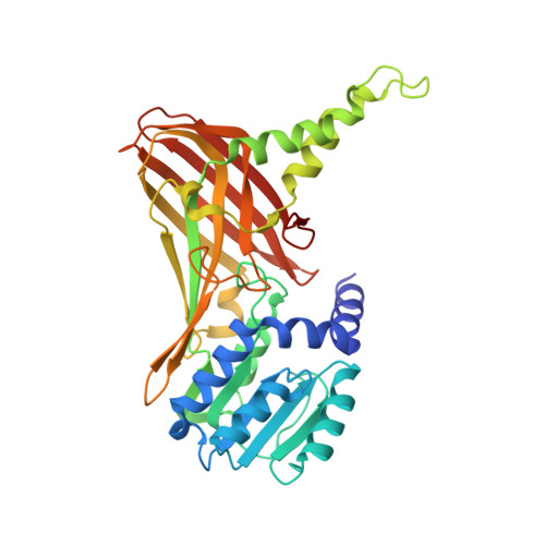
    Coactivator-associated arginine methyltransferase 1 (CARM1), a protein arginine methyltransferase recruited by several transcription factors, methylates a large variety of proteins and plays a critical role in gene expression. We report, in this paper, four crystal structures of isolated modules of CARM1. The 1.7 A crystal structure of the N-terminal domain of CARM1 reveals an unexpected PH domain, a scaffold frequently found to regulate protein-protein interactions in a large variety of biological processes. Three crystal structures of the CARM1 catalytic module, two free and one cofactor-bound forms (refined at 2.55 A, 2.4 A and 2.2 A, respectively) reveal large structural modifications including disorder to order transition, helix to strand transition and active site modifications. The N-terminal and the C-terminal end of CARM1 catalytic module contain molecular switches that may inspire how CARM1 regulates its biological activities by protein-protein interactions.


  • Organizational Affiliation
    • Département de Biologie et Génomique Structurales, IGBMC (Institut de Génétique et de Biologie Moléculaire et Cellulaire), UMR 7104 CNRS, U596 INSERM, ULP, Illkirch, France.

Macromolecule Content 

  • Total Structure Weight: 156.63 kDa 
  • Atom Count: 11,469 
  • Modeled Residue Count: 1,348 
  • Deposited Residue Count: 1,364 
  • Unique protein chains: 1

Macromolecules

Find similar proteins by:|  3D Structure
Entity ID: 1
MoleculeChains  Sequence LengthOrganismDetailsImage
Histone-arginine methyltransferase CARM1
A, B, C, D
341Rattus norvegicusMutation(s): 0 
Gene Names: Carm1Prmt4
EC: 2.1.1.125 (PDB Primary Data), 2.1.1 (PDB Primary Data), 2.1.1.319 (UniProt)
UniProt
Find proteins for Q4AE70 (Rattus norvegicus)
Explore Q4AE70 
Go to UniProtKB:  Q4AE70
Entity Groups
Sequence Clusters30% Identity50% Identity70% Identity90% Identity95% Identity100% Identity
UniProt GroupQ4AE70
Sequence Annotations
Expand
Reference Sequence

Experimental Data & Validation

Experimental Data

  • Method: X-RAY DIFFRACTION
  • Resolution: 2.20 Å
  • R-Value Free:  0.234 (Depositor), 0.231 (DCC) 
  • R-Value Work:  0.182 (Depositor), 0.181 (DCC) 
  • R-Value Observed: 0.185 (Depositor) 
Space Group: P 21 21 2
Unit Cell:
Length ( Å )Angle ( ˚ )
a = 74.819α = 90
b = 98.689β = 90
c = 206.926γ = 90
Software Package:
Software NamePurpose
REFMACrefinement
HKL-2000data collection
DENZOdata reduction
SCALEPACKdata scaling

Structure Validation

View Full Validation Report


Ligand Structure Quality Assessment 


Entry History 

Deposition Data

Revision History  (Full details and data files)

  • Version 1.0: 2007-11-06
    Type: Initial release
  • Version 1.1: 2011-07-13
    Changes: Advisory, Refinement description, Version format compliance
  • Version 1.2: 2024-02-21
    Changes: Data collection, Database references, Derived calculations