3B3B | pdb_00003b3b

Crystal structure of E. coli Aminopeptidase N in complex with tryptophan


Experimental Data Snapshot

  • Method: X-RAY DIFFRACTION
  • Resolution: 1.85 Å
  • R-Value Free: 
    0.178 (Depositor), 0.178 (DCC) 
  • R-Value Work: 
    0.151 (Depositor), 0.151 (DCC) 
  • R-Value Observed: 
    0.153 (Depositor) 

Starting Model: experimental
View more details

wwPDB Validation 3D Report Full Report

Validation slider image for 3B3B

Ligand Structure Quality Assessment 


This is version 1.2 of the entry. See complete history

Literature

Structural basis for the unusual specificity of Escherichia coli aminopeptidase N.

Addlagatta, A.Gay, L.Matthews, B.W.

(2008) Biochemistry 47: 5303-5311

  • DOI: https://doi.org/10.1021/bi7022333
  • Primary Citation Related Structures: 
    3B2P, 3B2X, 3B34, 3B37, 3B3B

  • PubMed Abstract: 

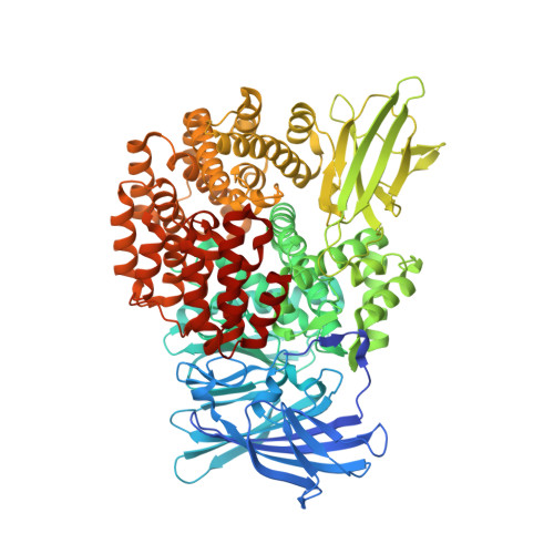
    Aminopeptidase N from Escherichia coli is a M1 class aminopeptidase with the active-site region related to that of thermolysin. The enzyme has unusual specificity, cleaving adjacent to the large, nonpolar amino acids Phe and Tyr but also cleaving next to the polar residues Lys and Arg. To try to understand the structural basis for this pattern of hydrolysis, the structure of the enzyme was determined in complex with the amino acids L-arginine, L-lysine, L-phenylalanine, L-tryptophan, and L-tyrosine. These amino acids all bind with their backbone atoms close to the active-site zinc ion and their side chain occupying the S1 subsite. This subsite is in the form of a cylinder, about 10 A in cross-section and 12 A in length. The bottom of the cylinder includes the zinc ion and a number of polar side chains that make multiple hydrogen-bonding and other interactions with the alpha-amino group and the alpha-carboxylate of the bound amino acid. The walls of the S1 cylinder are hydrophobic and accommodate the nonpolar or largely nonpolar side chains of Phe and Tyr. The top of the cylinder is polar in character and includes bound water molecules. The epsilon-amino group of the bound lysine side chain and the guanidinium group of arginine both make multiple hydrogen bonds to this part of the S1 site. At the same time, the hydrocarbon part of the lysine and arginine side chains is accommodated within the nonpolar walls of the S1 cylinder. This combination of hydrophobic and hydrophilic binding surfaces explains the ability of ePepN to cleave Lys, Arg, Phe, and Tyr. Another favored substrate has Ala at the P1 position. The short, nonpolar side chain of this residue can clearly be bound within the hydrophobic part of the S1 cylinder, but the reason for its facile hydrolysis remains uncertain.


  • Organizational Affiliation
    • Institute of Molecular Biology, Howard Hughes Medical Institute, and Department of Physics, 1229 University of Oregon, Eugene, Oregon 97403-1229, USA.

Macromolecule Content 

  • Total Structure Weight: 102.14 kDa 
  • Atom Count: 8,191 
  • Modeled Residue Count: 866 
  • Deposited Residue Count: 891 
  • Unique protein chains: 1

Macromolecules

Find similar proteins by:|  3D Structure
Entity ID: 1
MoleculeChains  Sequence LengthOrganismDetailsImage
Aminopeptidase N891Escherichia coli K-12Mutation(s): 0 
Gene Names: Aminopeptidase N
EC: 3.4.11.2
UniProt
Find proteins for P04825 (Escherichia coli (strain K12))
Explore P04825 
Go to UniProtKB:  P04825
Entity Groups
Sequence Clusters30% Identity50% Identity70% Identity90% Identity95% Identity100% Identity
UniProt GroupP04825
Sequence Annotations
Expand
Reference Sequence

Small Molecules

Ligands 5 Unique
IDChains Name / Formula / InChI Key2D Diagram3D Interactions
TRP

Query on TRP


Download:Ideal Coordinates CCD File
E [auth A]TRYPTOPHAN
C11 H12 N2 O2
QIVBCDIJIAJPQS-VIFPVBQESA-N
MLI

Query on MLI


Download:Ideal Coordinates CCD File
F [auth A]MALONATE ION
C3 H2 O4
OFOBLEOULBTSOW-UHFFFAOYSA-L
GOL

Query on GOL


Download:Ideal Coordinates CCD File
G [auth A],
H [auth A],
I [auth A]
GLYCEROL
C3 H8 O3
PEDCQBHIVMGVHV-UHFFFAOYSA-N
ZN

Query on ZN


Download:Ideal Coordinates CCD File
B [auth A]ZINC ION
Zn
PTFCDOFLOPIGGS-UHFFFAOYSA-N
NA

Query on NA


Download:Ideal Coordinates CCD File
C [auth A],
D [auth A]
SODIUM ION
Na
FKNQFGJONOIPTF-UHFFFAOYSA-N

Experimental Data & Validation

Experimental Data

  • Method: X-RAY DIFFRACTION
  • Resolution: 1.85 Å
  • R-Value Free:  0.178 (Depositor), 0.178 (DCC) 
  • R-Value Work:  0.151 (Depositor), 0.151 (DCC) 
  • R-Value Observed: 0.153 (Depositor) 
Space Group: P 31 2 1
Unit Cell:
Length ( Å )Angle ( ˚ )
a = 120.64α = 90
b = 120.64β = 90
c = 170.64γ = 120
Software Package:
Software NamePurpose
REFMACrefinement
PDB_EXTRACTdata extraction
BOSdata collection
HKL-2000data reduction
HKL-2000data scaling
MOLREPphasing

Structure Validation

View Full Validation Report


Ligand Structure Quality Assessment 


Entry History 

Deposition Data

Revision History  (Full details and data files)

  • Version 1.0: 2008-05-06
    Type: Initial release
  • Version 1.1: 2011-07-13
    Changes: Non-polymer description, Version format compliance
  • Version 1.2: 2023-08-30
    Changes: Data collection, Database references, Derived calculations, Refinement description