3B2D | pdb_00003b2d

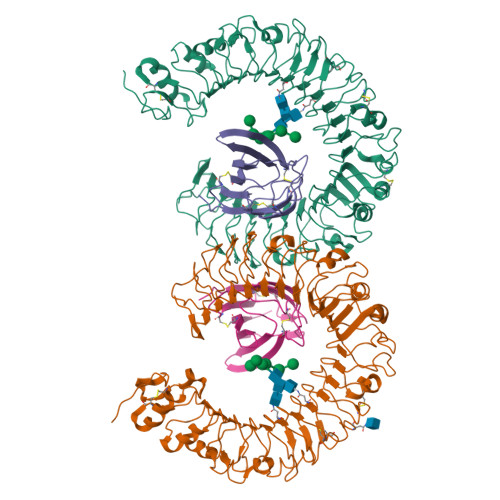
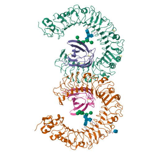
Crystal structure of human RP105/MD-1 complex


Experimental Data Snapshot

  • Method: X-RAY DIFFRACTION
  • Resolution: 2.80 Å
  • R-Value Free: 
    0.284 (Depositor), 0.284 (DCC) 
  • R-Value Work: 
    0.218 (Depositor), 0.218 (DCC) 
  • R-Value Observed: 
    0.221 (Depositor) 

wwPDB Validation   3D Report Full Report


Ligand Structure Quality Assessment 

Created with Raphaël 2.3.0Worse 01 BetterLigand structure goodness of fit to experimental dataBest fitted NAGClick on this verticalbar to view details

This is version 2.1 of the entry. See complete history


Literature

Crystal Structures of Mouse and Human RP105/MD-1 Complexes Reveal Unique Dimer Organization of the Toll-Like Receptor Family.

Ohto, U.Miyake, K.Shimizu, T.

(2011) J Mol Biology 413: 815-825

  • DOI: https://doi.org/10.1016/j.jmb.2011.09.020
  • Primary Citation of Related Structures:  
    3B2D, 3T6Q

  • PubMed Abstract: 

    The Toll-like receptor (TLR) 4/MD-2 heterodimer senses lipopolysaccharide (LPS). RP105 (radioprotective 105 kDa), a TLR-related molecule, is similar to TLR4 in that the extracellular leucine-rich repeats associate with MD-1, the MD-2-like molecule. MD-2 has a unique hydrophobic cavity that directly binds to lipid A, the active center of LPS. LPS-bound MD-2 opens the secondary interface with TLR4, leading to dimerization of TLR4/MD-2. MD-1 also has a hydrophobic cavity that accommodates lipid IVa, a precursor of lipid A, suggesting a role for the RP105/MD-1 heterodimer in sensing LPS or related microbial products. Little is known, however, about the structure of the RP105/MD-1 heterodimer or its oligomer. Here, we have determined the crystal structures of mouse and human RP105/MD-1 complexes at 1.9 and 2.8 Å resolutions, respectively. Both mouse and human RP105/MD-1 exhibit dimerization of the 1:1 RP105/MD-1 complex, demonstrating a novel organization. The "m"-shaped 2:2 RP105/MD-1 complex exhibits an inverse arrangement, with N-termini interacting in the middle. Thus, the dimerization interface of RP105/MD-1 is located on the opposite side of the complex, compared to the 2:2 TLR4/MD-2 complex. These results demonstrate that the 2:2 RP105/MD-1 complex is distinct from previously reported TLR dimers, including TLR4/MD-2, TLR1/TLR2, TLR2/TLR6, and TLR3, all of which facilitate homotypic or heterotypic interaction of the C-terminal cytoplasmic signaling domain.


  • Organizational Affiliation

    Graduate School of Pharmaceutical Sciences, University of Tokyo, Hongo, Bunkyo-ku, Tokyo 113-0033, Japan.


Macromolecules
Find similar proteins by:  (by identity cutoff)  |  3D Structure
Entity ID: 1
MoleculeChains Sequence LengthOrganismDetailsImage
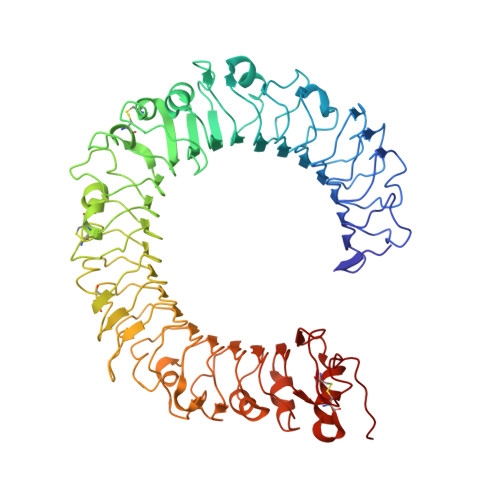
CD180 antigen
A, B
603Homo sapiensMutation(s): 0 
Gene Names: CD180LY64RP105
UniProt & NIH Common Fund Data Resources
Find proteins for Q99467 (Homo sapiens)
Explore Q99467 
Go to UniProtKB:  Q99467
PHAROS:  Q99467
GTEx:  ENSG00000134061 
Entity Groups  
Sequence Clusters30% Identity50% Identity70% Identity90% Identity95% Identity100% Identity
UniProt GroupQ99467
Glycosylation
Glycosylation Sites: 2Go to GlyGen: Q99467-1
Sequence Annotations
Expand
  • Reference Sequence
Find similar proteins by:  (by identity cutoff)  |  3D Structure
Entity ID: 2
MoleculeChains Sequence LengthOrganismDetailsImage
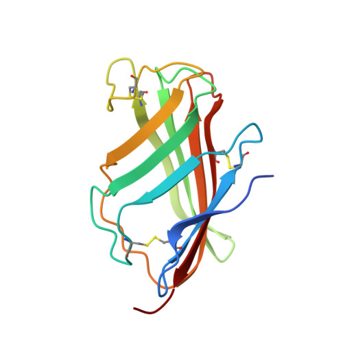
Lymphocyte antigen 86
C, D
144Homo sapiensMutation(s): 0 
Gene Names: LY86MD1
UniProt & NIH Common Fund Data Resources
Find proteins for O95711 (Homo sapiens)
Explore O95711 
Go to UniProtKB:  O95711
PHAROS:  O95711
GTEx:  ENSG00000112799 
Entity Groups  
Sequence Clusters30% Identity50% Identity70% Identity90% Identity95% Identity100% Identity
UniProt GroupO95711
Sequence Annotations
Expand
  • Reference Sequence
Oligosaccharides

Help

Entity ID: 3
MoleculeChains Length2D Diagram Glycosylation3D Interactions
alpha-D-mannopyranose-(1-2)-alpha-D-mannopyranose-(1-3)-[alpha-D-mannopyranose-(1-2)-alpha-D-mannopyranose-(1-6)]alpha-D-mannopyranose-(1-6)-[alpha-D-mannopyranose-(1-3)]beta-D-mannopyranose-(1-4)-2-acetamido-2-deoxy-beta-D-glucopyranose-(1-4)-2-acetamido-2-deoxy-beta-D-glucopyranose
E
9N-Glycosylation
Glycosylation Resources
GlyTouCan:  G37135JQ
GlyCosmos:  G37135JQ
GlyGen:  G37135JQ
Entity ID: 4
MoleculeChains Length2D Diagram Glycosylation3D Interactions
2-acetamido-2-deoxy-beta-D-glucopyranose-(1-4)-2-acetamido-2-deoxy-beta-D-glucopyranose
F, H
2N-Glycosylation
Glycosylation Resources
GlyTouCan:  G42666HT
GlyCosmos:  G42666HT
GlyGen:  G42666HT
Entity ID: 5
MoleculeChains Length2D Diagram Glycosylation3D Interactions
alpha-D-mannopyranose-(1-2)-alpha-D-mannopyranose-(1-6)-[alpha-D-mannopyranose-(1-3)]alpha-D-mannopyranose-(1-6)-[alpha-D-mannopyranose-(1-3)]beta-D-mannopyranose-(1-4)-2-acetamido-2-deoxy-beta-D-glucopyranose-(1-4)-2-acetamido-2-deoxy-beta-D-glucopyranose
G
8N-Glycosylation
Glycosylation Resources
GlyTouCan:  G61846BY
GlyCosmos:  G61846BY
GlyGen:  G61846BY
Small Molecules
Ligands 1 Unique
IDChains Name / Formula / InChI Key2D Diagram3D Interactions
NAG
Query on NAG

Download Ideal Coordinates CCD File 
I [auth B]2-acetamido-2-deoxy-beta-D-glucopyranose
C8 H15 N O6
OVRNDRQMDRJTHS-FMDGEEDCSA-N
Experimental Data & Validation

Experimental Data

  • Method: X-RAY DIFFRACTION
  • Resolution: 2.80 Å
  • R-Value Free:  0.284 (Depositor), 0.284 (DCC) 
  • R-Value Work:  0.218 (Depositor), 0.218 (DCC) 
  • R-Value Observed: 0.221 (Depositor) 
Space Group: P 21 21 2
Unit Cell:
Length ( Å )Angle ( ˚ )
a = 203.997α = 90
b = 66.441β = 90
c = 119.097γ = 90
Software Package:
Software NamePurpose
MOLREPphasing
REFMACrefinement
HKL-2000data reduction
HKL-2000data scaling

Structure Validation

View Full Validation Report


Ligand Structure Quality Assessment 

Created with Raphaël 2.3.0Worse 01 BetterLigand structure goodness of fit to experimental dataBest fitted NAGClick on this verticalbar to view details

Entry History 

Deposition Data

Revision History  (Full details and data files)

  • Version 1.0: 2011-11-09
    Type: Initial release
  • Version 2.0: 2020-07-29
    Type: Remediation
    Reason: Carbohydrate remediation
    Changes: Atomic model, Data collection, Database references, Derived calculations, Structure summary
  • Version 2.1: 2024-11-20
    Changes: Data collection, Database references, Structure summary