3B0O | pdb_00003b0o

Crystal structure of alpha-lactalbumin


Experimental Data Snapshot

  • Method: X-RAY DIFFRACTION
  • Resolution: 1.61 Å
  • R-Value Free: 
    0.193 (Depositor), 0.207 (DCC) 
  • R-Value Work: 
    0.153 (Depositor) 
  • R-Value Observed: 
    0.155 (Depositor) 

wwPDB Validation 3D Report Full Report

Validation slider image for 3B0O

This is version 1.2 of the entry. See complete history

Literature

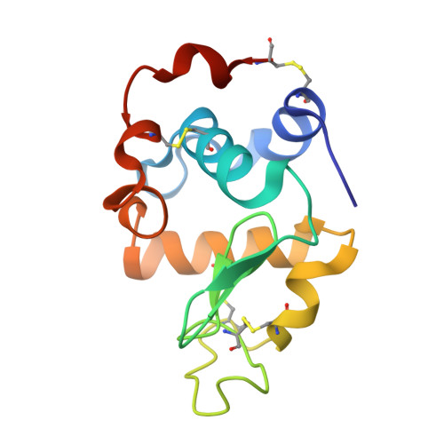
Structural insights into the stability perturbations induced by N-terminal variation in human and goat alpha-lactalbumin

Makabe, K.Nakamura, T.Kuwajima, K.

(2013) Protein Eng Des Sel 26: 165-170

  • DOI: https://doi.org/10.1093/protein/gzs093
  • Primary Citation Related Structures: 
    3B0I, 3B0K, 3B0O

  • PubMed Abstract: 

    Addition of an extra methionine at the N-terminus by recombinant expression of α-lactalbumin in Escherichia coli significantly destabilizes the protein, and this destabilization has hampered mutational analyses such as the mutational phi-value analysis of the protein. Deletion of residue 1 from the recombinant form recovers the stability in human and goat α-lactalbumin. Here, we thus determined the crystal structures of the residue 1-deletion variants of recombinant human and goat α-lactalbumin, and compared the structures with those of the authentic and recombinant forms. The results demonstrate the importance of the N-terminal backbone structure and hydrogen-bonding pattern for the stability of α-lactalbumin.


  • Organizational Affiliation
    • Okazaki Institute for Integrative Bioscience and Institute for Molecular Science, National Institutes of Natural Sciences, 5-1 Higashiyama, Myodaiji, Okazaki 444-8787, Japan. makabe@yz.yamagata-u.ac.jp

Macromolecule Content 

  • Total Structure Weight: 28.27 kDa 
  • Atom Count: 2,205 
  • Modeled Residue Count: 240 
  • Deposited Residue Count: 246 
  • Unique protein chains: 1

Macromolecules

Find similar proteins by:|  3D Structure
Entity ID: 1
MoleculeChains  Sequence LengthOrganismDetailsImage
Alpha-lactalbumin
A, B
123Homo sapiensMutation(s): 0 
Gene Names: LALBALYZL7
UniProt & NIH Common Fund Data Resources
Find proteins for P00709 (Homo sapiens)
Explore P00709 
Go to UniProtKB:  P00709
PHAROS:  P00709
GTEx:  ENSG00000167531 
Entity Groups
Sequence Clusters30% Identity50% Identity70% Identity90% Identity95% Identity100% Identity
UniProt GroupP00709
Sequence Annotations
Expand
Reference Sequence

Experimental Data & Validation

Experimental Data

  • Method: X-RAY DIFFRACTION
  • Resolution: 1.61 Å
  • R-Value Free:  0.193 (Depositor), 0.207 (DCC) 
  • R-Value Work:  0.153 (Depositor) 
  • R-Value Observed: 0.155 (Depositor) 
Space Group: P 1 21 1
Unit Cell:
Length ( Å )Angle ( ˚ )
a = 32.917α = 90
b = 68.161β = 92.22
c = 46.212γ = 90
Software Package:
Software NamePurpose
HKL-3000data collection
MOLREPphasing
REFMACrefinement
HKL-3000data reduction
HKL-3000data scaling

Structure Validation

View Full Validation Report


Entry History 

Deposition Data

Revision History  (Full details and data files)

  • Version 1.0: 2012-06-13
    Type: Initial release
  • Version 1.1: 2014-01-15
    Changes: Database references
  • Version 1.2: 2024-10-30
    Changes: Data collection, Database references, Derived calculations, Structure summary