3AX8

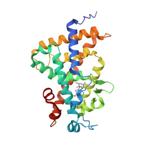
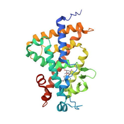
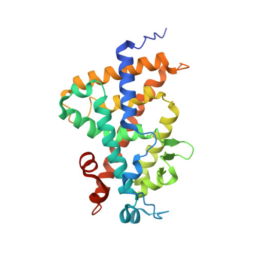
Crystal structure of the human vitamin D receptor ligand binding domain complexed with 15alpha-methoxy-1alpha,25-dihydroxyvitamin D3


Experimental Data Snapshot

  • Method: X-RAY DIFFRACTION
  • Resolution: 2.60 Å
  • R-Value Free: 0.294 
  • R-Value Work: 0.215 
  • R-Value Observed: 0.218 

Starting Model: experimental
View more details

wwPDB Validation   3D Report Full Report


Ligand Structure Quality Assessment 


This is version 1.2 of the entry. See complete history


Literature

New C15-substituted active vitamin D3

Shindo, K.Kumagai, G.Takano, M.Sawada, D.Saito, N.Saito, H.Kakuda, S.Takagi, K.Ochiai, E.Horie, K.Takimoto-Kamimura, M.Ishizuka, S.Takenouchi, K.Kittaka, A.

(2011) Org Lett 13: 2852-2855

  • DOI: https://doi.org/10.1021/ol200828s
  • Primary Citation of Related Structures:  
    3AX8

  • PubMed Abstract: 

    C15-Substituted 1α,25-dihydroxyvitamin D(3) analogs were synthesized for the first time to investigate the effects of the modified CD-ring on biological activity concerning the agonistic positioning of helix-3 and helix-12 of the vitamin D receptor (VDR). X-ray cocrystallographic analysis proved that 0.6 Å shifts of the CD-ring and shrinking of the side chain were necessary to maintain the position of the 25-hydroxy group for proper interaction with helix-12. The 15-hydroxy-16-ene derivative showed higher binding affinity for hVDR than the natural hormone.


  • Organizational Affiliation

    Faculty of Pharmaceutical Sciences, Teikyo University, Sagamihara, Kanagawa, Japan.


Macromolecules
Find similar proteins by:  (by identity cutoff)  |  3D Structure
Entity ID: 1
MoleculeChains Sequence LengthOrganismDetailsImage
Vitamin D3 receptor263Homo sapiensMutation(s): 0 
Gene Names: VDRNR1I1
UniProt & NIH Common Fund Data Resources
Find proteins for P11473 (Homo sapiens)
Explore P11473 
Go to UniProtKB:  P11473
PHAROS:  P11473
GTEx:  ENSG00000111424 
Entity Groups  
Sequence Clusters30% Identity50% Identity70% Identity90% Identity95% Identity100% Identity
UniProt GroupP11473
Sequence Annotations
Expand
  • Reference Sequence
Small Molecules
Ligands 1 Unique
IDChains Name / Formula / InChI Key2D Diagram3D Interactions
EIM
Query on EIM

Download Ideal Coordinates CCD File 
B [auth A](1R,3S,5Z)-5-[(2E)-2-[(1R,3S,3aS,7aR)-1-[(2R)-6-hydroxy-6-methyl-heptan-2-yl]-3-methoxy-7a-methyl-2,3,3a,5,6,7-hexahydro-1H-inden-4-ylidene]ethylidene]-4-methylidene-cyclohexane-1,3-diol
C28 H46 O4
MKYCMLKYANTZFR-GSVKQXOCSA-N
Experimental Data & Validation

Experimental Data

  • Method: X-RAY DIFFRACTION
  • Resolution: 2.60 Å
  • R-Value Free: 0.294 
  • R-Value Work: 0.215 
  • R-Value Observed: 0.218 
  • Space Group: P 21 21 21
Unit Cell:
Length ( Å )Angle ( ˚ )
a = 44.489α = 90
b = 51.585β = 90
c = 132.017γ = 90
Software Package:
Software NamePurpose
HKL-2000data collection
MOLREPphasing
REFMACrefinement
HKL-2000data reduction
HKL-2000data scaling

Structure Validation

View Full Validation Report


Ligand Structure Quality Assessment 


Entry History 

Deposition Data

Revision History  (Full details and data files)

  • Version 1.0: 2011-10-05
    Type: Initial release
  • Version 1.1: 2017-08-16
    Changes: Refinement description, Source and taxonomy
  • Version 1.2: 2023-11-01
    Changes: Data collection, Database references, Derived calculations, Refinement description