3AWM | pdb_00003awm

Cytochrome P450SP alpha (CYP152B1) wild-type with palmitic acid


Experimental Data Snapshot

  • Method: X-RAY DIFFRACTION
  • Resolution: 1.65 Å
  • R-Value Free: 
    0.173 (Depositor), 0.203 (DCC) 
  • R-Value Work: 
    0.151 (Depositor), 0.206 (DCC) 
  • R-Value Observed: 
    0.152 (Depositor) 

wwPDB Validation 3D Report Full Report

Validation slider image for 3AWM

Ligand Structure Quality Assessment 


This is version 1.3 of the entry. See complete history

Literature

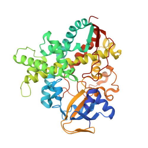
Crystal structure of H2O2-dependent cytochrome P450SPalpha with its bound fatty acid substrate: insight into the regioselective hydroxylation of fatty acids at the alpha position

Fujishiro, T.Shoji, O.Nagano, S.Sugimoto, H.Shiro, Y.Watanabe, Y.

(2011) J Biological Chem 286: 29941-29950

  • DOI: https://doi.org/10.1074/jbc.M111.245225
  • Primary Citation Related Structures: 
    3AWM, 3AWP, 3AWQ

  • PubMed Abstract: 

    Cytochrome P450(SPα) (CYP152B1) isolated from Sphingomonas paucimobilis is the first P450 to be classified as a H(2)O(2)-dependent P450. P450(SPα) hydroxylates fatty acids with high α-regioselectivity. Herein we report the crystal structure of P450(SPα) with palmitic acid as a substrate at a resolution of 1.65 Å. The structure revealed that the C(α) of the bound palmitic acid in one of the alternative conformations is 4.5 Å from the heme iron. This conformation explains the highly selective α-hydroxylation of fatty acid observed in P450(SPα). Mutations at the active site and the F-G loop of P450(SPα) did not impair its regioselectivity. The crystal structures of mutants (L78F and F288G) revealed that the location of the bound palmitic acid was essentially the same as that in the WT, although amino acids at the active site were replaced with the corresponding amino acids of cytochrome P450(BSβ) (CYP152A1), which shows β-regioselectivity. This implies that the high regioselectivity of P450(SPα) is caused by the orientation of the hydrophobic channel, which is more perpendicular to the heme plane than that of P450(BSβ).


  • Organizational Affiliation
    • Department of Chemistry, Graduate School of Science, Nagoya University, Furo-cho, Chikusa-ku, Nagoya, Japan.

Macromolecule Content 

  • Total Structure Weight: 47.54 kDa 
  • Atom Count: 3,749 
  • Modeled Residue Count: 407 
  • Deposited Residue Count: 415 
  • Unique protein chains: 1

Macromolecules

Find similar proteins by:|  3D Structure
Entity ID: 1
MoleculeChains  Sequence LengthOrganismDetailsImage
Fatty acid alpha-hydroxylase415Sphingomonas paucimobilisMutation(s): 0 
EC: 1.11.2.4
UniProt
Find proteins for O24782 (Sphingomonas paucimobilis)
Explore O24782 
Go to UniProtKB:  O24782
Entity Groups
Sequence Clusters30% Identity50% Identity70% Identity90% Identity95% Identity100% Identity
UniProt GroupO24782
Sequence Annotations
Expand
Reference Sequence

Experimental Data & Validation

Experimental Data

  • Method: X-RAY DIFFRACTION
  • Resolution: 1.65 Å
  • R-Value Free:  0.173 (Depositor), 0.203 (DCC) 
  • R-Value Work:  0.151 (Depositor), 0.206 (DCC) 
  • R-Value Observed: 0.152 (Depositor) 
Space Group: P 31 2 1
Unit Cell:
Length ( Å )Angle ( ˚ )
a = 94.44α = 90
b = 94.44β = 90
c = 113.553γ = 120
Software Package:
Software NamePurpose
DENZOdata reduction
SCALEPACKdata scaling
SHELXphasing
REFMACrefinement
PDB_EXTRACTdata extraction
HKL-2000data collection
HKL-2000data reduction
HKL-2000data scaling
SHELXDphasing

Structure Validation

View Full Validation Report


Ligand Structure Quality Assessment 


Entry History 

Deposition Data

Revision History  (Full details and data files)

  • Version 1.0: 2011-06-29
    Type: Initial release
  • Version 1.1: 2011-07-13
    Changes: Version format compliance
  • Version 1.2: 2013-06-05
    Changes: Database references
  • Version 1.3: 2024-03-13
    Changes: Data collection, Database references, Derived calculations