3AQ0 | pdb_00003aq0

Ligand-bound form of Arabidopsis medium/long-chain length prenyl pyrophosphate synthase (surface polar residue mutant)


Experimental Data Snapshot

  • Method: X-RAY DIFFRACTION
  • Resolution: 2.65 Å
  • R-Value Free: 
    0.274 (Depositor), 0.285 (DCC) 
  • R-Value Work: 
    0.222 (Depositor), 0.250 (DCC) 
  • R-Value Observed: 
    0.225 (Depositor) 

Starting Model: experimental
View more details

wwPDB Validation 3D Report Full Report

Validation slider image for 3AQ0

Ligand Structure Quality Assessment 


This is version 1.4 of the entry. See complete history

Literature

Structure and mechanism of an Arabidopsis medium/long-chain-length prenyl pyrophosphate synthase

Hsieh, F.-L.Chang, T.-H.Ko, T.-P.Wang, A.H.-J.

(2011) Plant Physiol 155: 1079-1090

  • DOI: https://doi.org/10.1104/pp.110.168799
  • Primary Citation Related Structures: 
    3APZ, 3AQ0

  • PubMed Abstract: 

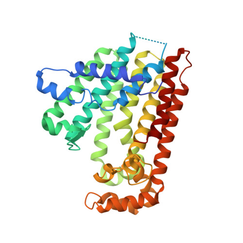
    Prenyltransferases (PTSs) are involved in the biosynthesis of terpenes with diverse functions. Here, a novel PTS from Arabidopsis (Arabidopsis thaliana) is identified as a trans-type polyprenyl pyrophosphate synthase (AtPPPS), which forms a trans-double bond during each homoallylic substrate condensation, rather than a homomeric C10-geranyl pyrophosphate synthase as originally proposed. Biochemical and genetic complementation analyses indicate that AtPPPS synthesizes C25 to C45 medium/long-chain products. Its close relationship to other long-chain PTSs is also uncovered by phylogenetic analysis. A mutant of contiguous surface polar residues was produced by replacing four charged surface amino acids with alanines to facilitate the crystallization of the enzyme. The crystal structures of AtPPPS determined here in apo and ligand-bound forms further reveal an active-site cavity sufficient to accommodate the medium/long-chain products. The two monomers in each dimer adopt different conformations at the entrance of the active site depending on the binding of substrates. Taken together, these results suggest that AtPPPS is endowed with a unique functionality among the known PTSs.


  • Organizational Affiliation
    • Institute of Biological Chemistry, Academia Sinica, Taipei 115, Taiwan.

Macromolecule Content 

  • Total Structure Weight: 307.05 kDa 
  • Atom Count: 20,426 
  • Modeled Residue Count: 2,551 
  • Deposited Residue Count: 2,784 
  • Unique protein chains: 1

Macromolecules

Find similar proteins by:|  3D Structure
Entity ID: 1
MoleculeChains  Sequence LengthOrganismDetailsImage
Geranyl diphosphate synthase
A, B, C, D, E
A, B, C, D, E, F, G, H
348Arabidopsis thalianaMutation(s): 4 
Gene Names: AtNPPPSgpps
EC: 2.5.1.30 (PDB Primary Data), 2.5.1.85 (UniProt)
UniProt
Find proteins for Q5HZ00 (Arabidopsis thaliana)
Explore Q5HZ00 
Go to UniProtKB:  Q5HZ00
Entity Groups
Sequence Clusters30% Identity50% Identity70% Identity90% Identity95% Identity100% Identity
UniProt GroupQ5HZ00
Sequence Annotations
Expand
Reference Sequence

Small Molecules

Ligands 5 Unique
IDChains Name / Formula / InChI Key2D Diagram3D Interactions
FPP

Query on FPP


Download:Ideal Coordinates CCD File
BA [auth G]
EA [auth H]
K [auth A]
N [auth B]
R [auth C]
BA [auth G],
EA [auth H],
K [auth A],
N [auth B],
R [auth C],
X [auth E]
FARNESYL DIPHOSPHATE
C15 H28 O7 P2
VWFJDQUYCIWHTN-YFVJMOTDSA-N
ISY

Query on ISY


Download:Ideal Coordinates CCD File
FA [auth H],
L [auth A]
3-methylbut-3-enylsulfanyl(phosphonooxy)phosphinic acid
C5 H12 O6 P2 S
YLTQZUVQWVAPNP-UHFFFAOYSA-N
PPV

Query on PPV


Download:Ideal Coordinates CCD File
AA [auth G]
M [auth B]
S [auth C]
T [auth D]
Y [auth E]
AA [auth G],
M [auth B],
S [auth C],
T [auth D],
Y [auth E],
Z [auth F]
PYROPHOSPHATE
H4 O7 P2
XPPKVPWEQAFLFU-UHFFFAOYSA-N
PEG

Query on PEG


Download:Ideal Coordinates CCD File
O [auth B],
U [auth D]
DI(HYDROXYETHYL)ETHER
C4 H10 O3
MTHSVFCYNBDYFN-UHFFFAOYSA-N
MG

Query on MG


Download:Ideal Coordinates CCD File
CA [auth H]
DA [auth H]
I [auth A]
J [auth A]
P [auth C]
CA [auth H],
DA [auth H],
I [auth A],
J [auth A],
P [auth C],
Q [auth C],
V [auth E],
W [auth E]
MAGNESIUM ION
Mg
JLVVSXFLKOJNIY-UHFFFAOYSA-N

Experimental Data & Validation

Experimental Data

  • Method: X-RAY DIFFRACTION
  • Resolution: 2.65 Å
  • R-Value Free:  0.274 (Depositor), 0.285 (DCC) 
  • R-Value Work:  0.222 (Depositor), 0.250 (DCC) 
  • R-Value Observed: 0.225 (Depositor) 
Space Group: P 61
Unit Cell:
Length ( Å )Angle ( ˚ )
a = 115.961α = 90
b = 115.961β = 90
c = 385.874γ = 120
Software Package:
Software NamePurpose
PHASERphasing
REFMACrefinement
PDB_EXTRACTdata extraction
HKL-2000data collection
DENZOdata reduction
SCALEPACKdata scaling
HKL-2000data scaling

Structure Validation

View Full Validation Report


Ligand Structure Quality Assessment 


Entry History 

Deposition Data

Revision History  (Full details and data files)

  • Version 1.0: 2011-01-19
    Type: Initial release
  • Version 1.1: 2011-07-13
    Changes: Version format compliance
  • Version 1.2: 2014-01-29
    Changes: Database references
  • Version 1.3: 2017-10-11
    Changes: Refinement description
  • Version 1.4: 2023-11-01
    Changes: Data collection, Database references, Derived calculations, Refinement description