3AO9 | pdb_00003ao9

Crystal structure of the C-terminal domain of sequence-specific ribonuclease


Experimental Data Snapshot

  • Method: X-RAY DIFFRACTION
  • Resolution: 2.10 Å
  • R-Value Free: 
    0.259 (Depositor), 0.258 (DCC) 
  • R-Value Work: 
    0.222 (Depositor), 0.224 (DCC) 
  • R-Value Observed: 
    0.224 (Depositor) 

Starting Model: experimental
View more details

wwPDB Validation 3D Report Full Report

Validation slider image for 3AO9

This is version 1.3 of the entry. See complete history

Literature

Identification of the catalytic residues of sequence-specific and histidine-free ribonuclease colicin E5

Inoue-Ito, S.Yajima, S.Fushinobu, S.Nakamura, S.Ogawa, T.Hidaka, M.Masaki, H.

(2012) J Biochem 152: 365-372

  • DOI: https://doi.org/10.1093/jb/mvs077
  • Primary Citation Related Structures: 
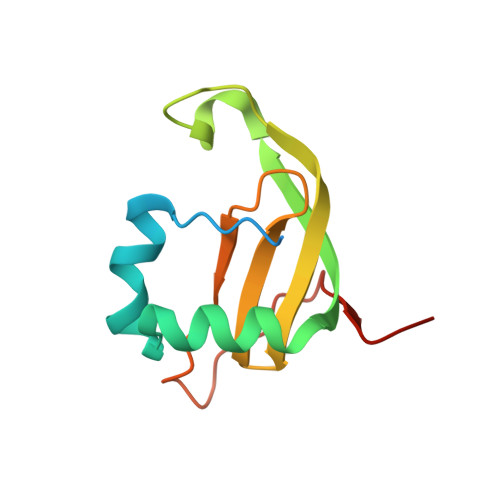
    3AO9, 3VJ7

  • PubMed Abstract: 

    Colicin E5 cleaves tRNAs for Tyr, His, Asn and Asp in their anticodons to abolish protein synthesis in Escherichia coli. We previously showed how its C-terminal RNase domain, E5-CRD, recognizes the anticodon bases but the catalytic mechanism remained to be elucidated. Although the reaction products with 5'-OH and 2',3'-cyclic phosphate ends suggested a similar mechanism to those of RNases A and T1, E5-CRD does not have the His residues necessary as a catalyst in usual RNases. To identify residues important for the catalytic reaction, mutants as to all residues within 5 Å from the central phosphorus of the scissile phosphodiester bond were prepared. Evaluation of the killing activities of the mutant colicins and the RNase activities of the mutant E5-CRDs suggested direct involvement of Arg33, Lys25, Gln29 and Lys60 in the reaction. Particularly, Arg33 plays a critical role and Ile94 provides a structural support of Arg33. Crystal structure of the complex of E5-CRD(R33Q)/dGpdUp showed structural and binding functional integrity of this mutant protein, suggesting involvement of Arg33 in the catalytic reaction. The structure of the free E5-CRD, we also determined, showed great flexibility of a flap region, which facilitates the access of Lys60 to the substrate in an induced-fit manner.


  • Organizational Affiliation
    • Department of Biotechnology, The University of Tokyo, Yayoi 1-1-1, Tokyo 113-8654, Japan.

Macromolecule Content 

  • Total Structure Weight: 26.16 kDa 
  • Atom Count: 1,547 
  • Modeled Residue Count: 183 
  • Deposited Residue Count: 232 
  • Unique protein chains: 1

Macromolecules

Find similar proteins by:|  3D Structure
Entity ID: 1
MoleculeChains  Sequence LengthOrganismDetailsImage
Colicin-E5
A, B
116Escherichia coliMutation(s): 0 
Gene Names: col
EC: 3.1
UniProt
Find proteins for P18000 (Escherichia coli)
Explore P18000 
Go to UniProtKB:  P18000
Entity Groups
Sequence Clusters30% Identity50% Identity70% Identity90% Identity95% Identity100% Identity
UniProt GroupP18000
Sequence Annotations
Expand
Reference Sequence

Experimental Data & Validation

Experimental Data

  • Method: X-RAY DIFFRACTION
  • Resolution: 2.10 Å
  • R-Value Free:  0.259 (Depositor), 0.258 (DCC) 
  • R-Value Work:  0.222 (Depositor), 0.224 (DCC) 
  • R-Value Observed: 0.224 (Depositor) 
Space Group: P 31 2 1
Unit Cell:
Length ( Å )Angle ( ˚ )
a = 62.71α = 90
b = 62.71β = 90
c = 100.51γ = 120
Software Package:
Software NamePurpose
ADSCdata collection
MOLREPphasing
REFMACrefinement
HKL-2000data reduction
HKL-2000data scaling

Structure Validation

View Full Validation Report


Entry History 

Deposition Data

Revision History  (Full details and data files)

  • Version 1.0: 2011-09-28
    Type: Initial release
  • Version 1.1: 2013-02-06
    Changes: Database references
  • Version 1.2: 2019-01-23
    Changes: Data collection, Structure summary
  • Version 1.3: 2023-11-01
    Changes: Data collection, Database references, Derived calculations, Refinement description